US5001225A - Monoclonal antibodies to a pan-malarial antigen - Google Patents

Monoclonal antibodies to a pan-malarial antigen Download PDF

Info

Publication number
US5001225A
US5001225A US06/939,113 US93911386A US5001225A US 5001225 A US5001225 A US 5001225A US 93911386 A US93911386 A US 93911386A US 5001225 A US5001225 A US 5001225A
Authority
US
United States
Prior art keywords
mab
antigen
antibody
present
monoclonal antibody
Prior art date
Legal status (The legal status is an assumption and is not a legal conclusion. Google has not performed a legal analysis and makes no representation as to the accuracy of the status listed.)
Expired - Fee Related
Application number
US06/939,113
Inventor
Diane W. Taylor
Current Assignee (The listed assignees may be inaccurate. Google has not performed a legal analysis and makes no representation or warranty as to the accuracy of the list.)
Georgetown University
Original Assignee
Georgetown University
Priority date (The priority date is an assumption and is not a legal conclusion. Google has not performed a legal analysis and makes no representation as to the accuracy of the date listed.)
Filing date
Publication date
Application filed by Georgetown University filed Critical Georgetown University
Priority to US06/939,113 priority Critical patent/US5001225A/en
Assigned to GEORGETOWN UNIVERSITY reassignment GEORGETOWN UNIVERSITY ASSIGNMENT OF ASSIGNORS INTEREST. Assignors: TAYLOR, DIANE W.
Priority to PCT/US1987/003252 priority patent/WO1988004302A1/en
Priority to AU10854/88A priority patent/AU1085488A/en
Application granted granted Critical
Publication of US5001225A publication Critical patent/US5001225A/en
Anticipated expiration legal-status Critical
Assigned to NATIONAL INSTITUTES OF HEALTH (NIH), U.S. DEPT. OF HEALTH AND HUMAN SERVICES (DHHS), U.S. GOVERNMENT reassignment NATIONAL INSTITUTES OF HEALTH (NIH), U.S. DEPT. OF HEALTH AND HUMAN SERVICES (DHHS), U.S. GOVERNMENT EXECUTIVE ORDER 9424, CONFIRMATORY LICENSE Assignors: GEORGETOWN UNIVERSITY
Expired - Fee Related legal-status Critical Current

Links

Images

Classifications

    • CCHEMISTRY; METALLURGY
    • C07ORGANIC CHEMISTRY
    • C07KPEPTIDES
    • C07K16/00Immunoglobulins [IGs], e.g. monoclonal or polyclonal antibodies
    • C07K16/18Immunoglobulins [IGs], e.g. monoclonal or polyclonal antibodies against material from animals or humans
    • C07K16/20Immunoglobulins [IGs], e.g. monoclonal or polyclonal antibodies against material from animals or humans from protozoa
    • C07K16/205Plasmodium
    • AHUMAN NECESSITIES
    • A61MEDICAL OR VETERINARY SCIENCE; HYGIENE
    • A61KPREPARATIONS FOR MEDICAL, DENTAL OR TOILETRY PURPOSES
    • A61K47/00Medicinal preparations characterised by the non-active ingredients used, e.g. carriers or inert additives; Targeting or modifying agents chemically bound to the active ingredient
    • A61K47/50Medicinal preparations characterised by the non-active ingredients used, e.g. carriers or inert additives; Targeting or modifying agents chemically bound to the active ingredient the non-active ingredient being chemically bound to the active ingredient, e.g. polymer-drug conjugates
    • A61K47/51Medicinal preparations characterised by the non-active ingredients used, e.g. carriers or inert additives; Targeting or modifying agents chemically bound to the active ingredient the non-active ingredient being chemically bound to the active ingredient, e.g. polymer-drug conjugates the non-active ingredient being a modifying agent
    • A61K47/68Medicinal preparations characterised by the non-active ingredients used, e.g. carriers or inert additives; Targeting or modifying agents chemically bound to the active ingredient the non-active ingredient being chemically bound to the active ingredient, e.g. polymer-drug conjugates the non-active ingredient being a modifying agent the modifying agent being an antibody, an immunoglobulin or a fragment thereof, e.g. an Fc-fragment
    • A61K47/6835Medicinal preparations characterised by the non-active ingredients used, e.g. carriers or inert additives; Targeting or modifying agents chemically bound to the active ingredient the non-active ingredient being chemically bound to the active ingredient, e.g. polymer-drug conjugates the non-active ingredient being a modifying agent the modifying agent being an antibody, an immunoglobulin or a fragment thereof, e.g. an Fc-fragment the modifying agent being an antibody or an immunoglobulin bearing at least one antigen-binding site
    • A61K47/6873Medicinal preparations characterised by the non-active ingredients used, e.g. carriers or inert additives; Targeting or modifying agents chemically bound to the active ingredient the non-active ingredient being chemically bound to the active ingredient, e.g. polymer-drug conjugates the non-active ingredient being a modifying agent the modifying agent being an antibody, an immunoglobulin or a fragment thereof, e.g. an Fc-fragment the modifying agent being an antibody or an immunoglobulin bearing at least one antigen-binding site the antibody targeting an immunoglobulin; the antibody being an anti-idiotypic antibody
    • CCHEMISTRY; METALLURGY
    • C07ORGANIC CHEMISTRY
    • C07KPEPTIDES
    • C07K14/00Peptides having more than 20 amino acids; Gastrins; Somatostatins; Melanotropins; Derivatives thereof
    • C07K14/435Peptides having more than 20 amino acids; Gastrins; Somatostatins; Melanotropins; Derivatives thereof from animals; from humans
    • C07K14/44Peptides having more than 20 amino acids; Gastrins; Somatostatins; Melanotropins; Derivatives thereof from animals; from humans from protozoa
    • C07K14/445Plasmodium
    • CCHEMISTRY; METALLURGY
    • C07ORGANIC CHEMISTRY
    • C07KPEPTIDES
    • C07K16/00Immunoglobulins [IGs], e.g. monoclonal or polyclonal antibodies
    • C07K16/42Immunoglobulins [IGs], e.g. monoclonal or polyclonal antibodies against immunoglobulins
    • C07K16/4208Immunoglobulins [IGs], e.g. monoclonal or polyclonal antibodies against immunoglobulins against an idiotypic determinant on Ig
    • C07K16/4241Immunoglobulins [IGs], e.g. monoclonal or polyclonal antibodies against immunoglobulins against an idiotypic determinant on Ig against anti-human or anti-animal Ig
    • C07K16/425Immunoglobulins [IGs], e.g. monoclonal or polyclonal antibodies against immunoglobulins against an idiotypic determinant on Ig against anti-human or anti-animal Ig against anti-protozoal Ig
    • GPHYSICS
    • G01MEASURING; TESTING
    • G01NINVESTIGATING OR ANALYSING MATERIALS BY DETERMINING THEIR CHEMICAL OR PHYSICAL PROPERTIES
    • G01N33/00Investigating or analysing materials by specific methods not covered by groups G01N1/00 - G01N31/00
    • G01N33/48Biological material, e.g. blood, urine; Haemocytometers
    • G01N33/50Chemical analysis of biological material, e.g. blood, urine; Testing involving biospecific ligand binding methods; Immunological testing
    • G01N33/53Immunoassay; Biospecific binding assay; Materials therefor
    • G01N33/569Immunoassay; Biospecific binding assay; Materials therefor for microorganisms, e.g. protozoa, bacteria, viruses
    • G01N33/56905Protozoa
    • GPHYSICS
    • G01MEASURING; TESTING
    • G01NINVESTIGATING OR ANALYSING MATERIALS BY DETERMINING THEIR CHEMICAL OR PHYSICAL PROPERTIES
    • G01N33/00Investigating or analysing materials by specific methods not covered by groups G01N1/00 - G01N31/00
    • G01N33/48Biological material, e.g. blood, urine; Haemocytometers
    • G01N33/50Chemical analysis of biological material, e.g. blood, urine; Testing involving biospecific ligand binding methods; Immunological testing
    • G01N33/68Chemical analysis of biological material, e.g. blood, urine; Testing involving biospecific ligand binding methods; Immunological testing involving proteins, peptides or amino acids
    • G01N33/6854Immunoglobulins
    • G01N33/686Anti-idiotype
    • AHUMAN NECESSITIES
    • A61MEDICAL OR VETERINARY SCIENCE; HYGIENE
    • A61KPREPARATIONS FOR MEDICAL, DENTAL OR TOILETRY PURPOSES
    • A61K38/00Medicinal preparations containing peptides
    • AHUMAN NECESSITIES
    • A61MEDICAL OR VETERINARY SCIENCE; HYGIENE
    • A61KPREPARATIONS FOR MEDICAL, DENTAL OR TOILETRY PURPOSES
    • A61K39/00Medicinal preparations containing antigens or antibodies
    • YGENERAL TAGGING OF NEW TECHNOLOGICAL DEVELOPMENTS; GENERAL TAGGING OF CROSS-SECTIONAL TECHNOLOGIES SPANNING OVER SEVERAL SECTIONS OF THE IPC; TECHNICAL SUBJECTS COVERED BY FORMER USPC CROSS-REFERENCE ART COLLECTIONS [XRACs] AND DIGESTS
    • Y02TECHNOLOGIES OR APPLICATIONS FOR MITIGATION OR ADAPTATION AGAINST CLIMATE CHANGE
    • Y02ATECHNOLOGIES FOR ADAPTATION TO CLIMATE CHANGE
    • Y02A50/00TECHNOLOGIES FOR ADAPTATION TO CLIMATE CHANGE in human health protection, e.g. against extreme weather
    • Y02A50/30Against vector-borne diseases, e.g. mosquito-borne, fly-borne, tick-borne or waterborne diseases whose impact is exacerbated by climate change
    • YGENERAL TAGGING OF NEW TECHNOLOGICAL DEVELOPMENTS; GENERAL TAGGING OF CROSS-SECTIONAL TECHNOLOGIES SPANNING OVER SEVERAL SECTIONS OF THE IPC; TECHNICAL SUBJECTS COVERED BY FORMER USPC CROSS-REFERENCE ART COLLECTIONS [XRACs] AND DIGESTS
    • Y10TECHNICAL SUBJECTS COVERED BY FORMER USPC
    • Y10STECHNICAL SUBJECTS COVERED BY FORMER USPC CROSS-REFERENCE ART COLLECTIONS [XRACs] AND DIGESTS
    • Y10S530/00Chemistry: natural resins or derivatives; peptides or proteins; lignins or reaction products thereof
    • Y10S530/808Materials and products related to genetic engineering or hybrid or fused cell technology, e.g. hybridoma, monoclonal products
    • Y10S530/809Fused cells, e.g. hybridoma
    • YGENERAL TAGGING OF NEW TECHNOLOGICAL DEVELOPMENTS; GENERAL TAGGING OF CROSS-SECTIONAL TECHNOLOGIES SPANNING OVER SEVERAL SECTIONS OF THE IPC; TECHNICAL SUBJECTS COVERED BY FORMER USPC CROSS-REFERENCE ART COLLECTIONS [XRACs] AND DIGESTS
    • Y10TECHNICAL SUBJECTS COVERED BY FORMER USPC
    • Y10STECHNICAL SUBJECTS COVERED BY FORMER USPC CROSS-REFERENCE ART COLLECTIONS [XRACs] AND DIGESTS
    • Y10S530/00Chemistry: natural resins or derivatives; peptides or proteins; lignins or reaction products thereof
    • Y10S530/863Chemistry: natural resins or derivatives; peptides or proteins; lignins or reaction products thereof involving IgM
    • Y10S530/864Monoclonal

Definitions

  • the present invention relates to a hybridoma cell line, designated 7H8, which secretes an IgM monoclonal antibody (MAb), designated MAb 7H8.
  • MAb 7H8 binds to an antigen associated with organisms of the genus Plasmodium.
  • the present invention is further directed to the discovery that MAb 7H8 is capable of binding to a novel antigen, designated Pf93, associated with P. falciparum.
  • the present invention is further directed to anti-idiotypic antibodies and to anti anti-idiotypic antibodies directed against MAb 7H8.
  • the present invention further relates to methods of detecting plasmodial infections in animals including humans employing MAb 7H8, to methods of suppressing plasmodial infections in animals, and to vaccines for and methods of vaccinating against P. falciparum.
  • Malaria a protozoan disease transmitted to humans by the Anopheles mosquito, is a serious infectious disease endemic to many parts of the world, especially tropical and subtropical regions. The disease is characterized by fever, rigors, anemia, splenomegaly, and chronic relapsing course.
  • Malaria is caused by protozoan organisms of the genus Plasmodium. Humans are infected by at least four species: P. vivax, P. ovale, P. malariae, and P. falciparum.
  • a female anopheline mosquito inoculates plasmodial sporozoites into the lymphohematogenous system while feeding. Following a brief stay in the peripheral blood system, the organisms invade hepatocytes, where they initiate the preclinical hepatic (exoerythrocytic) phase of the disease.
  • a single sporozoite can produce 2,000 to 40,000 hepatic merozoites by a process of asexual multiplication known as schizogony.
  • the daughter cells thus produced rupture back into the circulatory system in one to six weeks. This marks the end of the preclinical hepatic stage in some plasmodial species (P. falciparum and P. malariae), but other plasmodial species have persistent hepatic phases which produce episodes of bloodstream invasion for months and even years.
  • the clinical (erythrocytic) phase of malaria begins with the attachment of a released merozoite to the surface of a red blood cell.
  • the merozoite attaches at a specific receptor site, which varies for each malarial species. After attachment, the merozoite slowly works its way into the interior of the red blood cell. Once inside, the parasite appears as a ring-shaped trophozoite. The trophozoite later enlarges and becomes ameboid in appearance. The nucleus of the trophozoite later divides into several portions forming a multinucleated schizont. Cell cytoplasm slowly condenses around each daughter nucleus forming a new generation of merozoites. The invaded erythrocyte ruptures 48 to 72 hours later, releasing this new generation of merozoites, each of which can initiate a new red blood cell cycle.
  • red blood cells become filled with sexual forms of the parasite (gametocytes).
  • Gametocytes do not induce cell lysis, and will not develop further unless ingested by an Anopheles mosquito during a blood meal. Fertilization occurs in the stomach of the mosquito, and the resulting ookinete later forms a cyst on the outer surface of the mosquito's stomach, releasing many sporozoites. These sporozoites migrate to the mosquito's salivary glands and are inoculated into a human at the next blood meal, thus completing the cycle.
  • the most common diagnostic test for malaria is examination of an appropriately stained blood film. Morphological differences among the plasmodial species infecting humans allow identification in blood smears. Usually, fingertip blood is smeared on a glass slide and stained with Wright's, Giemsa's, or Field stain. Thick smears have the advantage of concentrating parasites. However, numerous artifacts will be found in such smears, with the result that a great deal of experience is required to correctly interpret such preparations.
  • Thin smears are easier to examine and have fewer artifacts. At the same time, the relative paucity of cells in a thin smear increases the likelihood that a diagnosis will be missed. Additionally, because the intensity of parasitemia varies greatly from hour to hour, frequent repeated examinations of blood smears may be required (e.g., every eight hours for two-three days), which may be prohibitively labor-intensive, especially in endemic areas.
  • the etiology, epidemiology, pathology, diagnosis, treatment, and prevention of malaria are described in Plorde, J.J., "Malaria", in, R.G. Petersdorf et al. eds. Harrison's Principles of Internal Medicine, Tenth Edition, McGraw-Hill Co., Publisher, New York, pp. 1187-1193 (1983).
  • antiplasmodial antibodies are produced during malarial infection in animals.
  • MAbs monoclonal antibodies
  • Plasmodium voelii a rodent malarial parasite.
  • MAbs identified monoclonal antibodies (MAbs) to Plasmodium voelii, a rodent malarial parasite.
  • MAbs identified stage-specific, species-specific, and cross-reactive antigens. The antigens were found on the surface or within the cytoplasm of the parasite, but not on the surface of erythrocytes from infected animals.
  • Malarial antigens are also known to appear in the serum of humans infected with P. falciparum. McGregor, I.A., et al., Lancet 1:881-884 (1968). The antigens may be secreted from infected erythrocytes or released during erythrocyte rupture and reinvasion. Winchell, E.J., et al., Molecular Biochemistry and Parasitology 10:287-296 (1984). Some circulating malarial antigens are biochemically stable to heating in boiling water for five minutes, and are thus termed heat-stable (S) antigens. Wilson, R.J.M., et al., Lancet 11:201-205 (1969). S antigens from various isolates of P.
  • S heat-stable
  • circulating antigens may form immune complexes and may thus play an important role in the pathogenesis of malaria, especially glomerulonephritis. Houba, V., Bull. W. H. O. 52:199-207 (1975).
  • Such a serodiagnostic method preferably would employ a monoclonal antibody specific for plasmodium-associated antigen. If an antibody were found that recognized an antigen common to all plasmodial species, this would allow the convenient identification of plasmodial infection, as, for example, in an initial screening.
  • the present inventor succeeded in isolating a hybridoma cell line, designated cell line 7H8, which secretes an IgM monoclonal antibody, designated MAb 7H8, which binds to a protein (antigen) present in organisms of the genus Plasmodium.
  • MAb 7H8 thus identifies a pan-species malarial antigen, and is suitable for use in immunoassays.
  • the present inventor has developed a two-sited assay with MAb 7H8 which takes advantage of the discovery that the 7H8 antigen has at least two epitopes per molecule.
  • MAb 7H8 recognizes a unique antigen (Pf93) found only in P. falciparum. MAb 7H8 thus may be used to isolate P. falciparum antigen Pf93, as well as for identification of general plasmodial infection.
  • Anti-idiotypic and anti anti-idiotypic antibodies have also been developed according to the present invention. The antigens and antibodies of the present invention are useful in immunodiagnostic and immunotherapeutic methods for the detection and treatment of plasmodial infections in animals, including humans.
  • FIG. 1 IIF patterns. P. voelii (17XL) parasites were treated with (A) normal mouse serum, (B) polyspecific immune serum, and (C) MAb 7H8. MAb 7H8 produced a similar pattern of fluorescence when assayed on (D) 17XNL P. voelii, (E) P. berghei, and (F) P. falciparum. Bar, 3 um.
  • FIG. 2 Molecular weight determinations for antigens bound by MAb 7H8. Immune precipitation results were obtained when an extract of [ 35 S]methionine-labeled 17XL P. voelii parasites was immune-precipitated with normal mouse serum (NMS) (lane 1), immune serum (IS) (lane 2), MAb 8B11 to a P. voelii antigen of 230 kDa (lane 3), and MAb 7H8 (lane 4). Immune precipitates were assayed on a 10% SDS-PAGE under nonreducing conditions. Western blot analysis: extracts of either normal mouse erythrocytes (E), P. berghei (Pb)-, or P.
  • E normal mouse erythrocytes
  • Pb P. berghei
  • voelii (Py)-infected erythrocytes were separated on a 5 to 15% SDS-PAGE under reducing conditions and then electrophoresed onto nitrocellulose paper. Samples were treated with 8B11, an IgM MAb to a 230,000-kDa protein of P. voelii known to cross-react with P. berghei and MAb 7H8.
  • FIG. 3 Direct binding of alkaline-phosphatase (AP)-labeled MAb 7H8 to three extracts of P. voelii.
  • Wells were coated with 100 ul of 1 mg of water-soluble parasite cytosol. Triton-X-100-extracted membrane proteins, or Triton-X-100-insoluble erythrocyte-parasite cytoskeletons per ml (the suspension was prepared in a tissue homogenizer before use).
  • Antigen-coated wells treated with PBS served as the blank.
  • the alkaline-phosphatase-labeled conjugate was used at a 1:500 dilution.
  • FIG. 4 Use of a two-sited assay for the detection of Ag-7H8 in extracts of P. voelii cytosol and plasma samples from mice with acute malaria.
  • the upper portion shows that, in general, Ag-7H8 was captured by wells coated with MAb 7H8 but not with BSA. The exception was P. voelii extract 2.
  • the lower portion demonstrates the heat-stable nature of Ag-7H8.
  • the four antigen extracts on the left contained cytosol isolated from approximately (1) 10 7 , (2) 2.5 ⁇ 10 7 , (3) 10 6 , and (4) 10 6 Percoll-isolated, infected erythrocytes.
  • Example 1 The acute-phase plasma samples on the right were collected on days 5 (samples 1 and 5) and 6 (samples 2 and 3) after infection with 17XL P. voelii and on day 7 (sample 4) after inoculation with 17XNL P. voelii.Samples contained plasma pooled from 20 to 50 BALB/c mice.
  • FIG. 5 Determination of the optimal amount of partially purified MAb 7H8 for application to microtiter wells.
  • step 1 microtiter wells were coated with various amounts of from 1 to 1,000 ug of partially purified MAb 7H8 per ml.
  • PBS, normal mouse serum (NMS), acute-phase plasma samples from mice with 30% parasitemia, and P. voelii cytosol extract (Ag) ( 1 mg/ml) were diluted 1:1 with 3% BSA in PBS and used in step 2.
  • a 1:500 dilution of alkaline-phosphatase-labeled MAb 7H8 was used in step 3. Results are expressed as means ⁇ 1 standard deviation for triplicate samples.
  • FIG. 6 Detection of Ag-7H8 in the serum of mice during 17XL P. voelii infection.
  • Groups of 21 and 15 BALB/c mice were infected with 10 4 and 10 6 17 XL P. voelii parasites, respectively.
  • Three mice in each group were bled out daily, and plasma samples from individual mice were pooled and examined in the two-sited assay.
  • the left axis and ascending curve show the course of parasitemia (the mean ⁇ the standard error of the mean for three mice).
  • the right axis and bars show results of the two-sited assay. Results are expressed as means ⁇ 1 standard deviation for triplicate samples. Normal BALB/c plasma samples (-------) were used as a control.
  • FIG. 7 Detection of Ag-7H8 in the plasma samples of various strains of mice infected with 17XNL strain of P. voelii. Groups of nine mice each were infected with 10 6 17XNL P. voelii parasites. Parasitemias (the left axis and ascending curves) are the mean for six to nine animals. Blood samples, collected from three mice on the days indicated, were pooled and assayed in the two-sited assay. Results (the right axis and bars) are expressed as means ⁇ 1 standard deviation for triplicate samples. Results for all C57B1/6 (B6) congenics were virtually identical, so only one is shown.
  • FIG. 8 Indirect Immunofluorescence pattern produced by normal mouse serum (negative control), immune serum on rings, trophozoites and schizonts, and MAb 7H8 on P. voelii and P. falciparum.
  • FIG. 9 Western blot analysis of antigen 7H8 in P. berghei (Pb), P. voelli (Py), and P. falciparum (Pf).
  • FIG. 10 Schematic diagram showing production of a portion of antigen Pf93 in the lambda gt11/E. coli Y1090 system.
  • FIG. 11 Flow diagram showing steps of production and release of Pf93 in P. falciparum.
  • FIG. 12 IFA analysis of course of Pf93 production after RBC infection.
  • FIG. 13 Schematic diagram showing two-sited assay using MAb 7H8.
  • FIG. 14 Time course of antigen release from infected RBC's determined by two-sited assay using MAb 7H8.
  • FIG. 15 Antigen release following reinvasion of RBC's determined by two-sited assay using MAb 7H8.
  • FIG. 16 Detection by two-sited assay using MAb 7H8 of antigen Pf93 in sera from individuals uninfected and acutely infected with P. falciparum.
  • FIG. 17 In vitro inhibition of merozoite invasion in human erythrocytes by MAb 7H8.
  • FIG. 18 Graph showing reduced parasitemia in 17XNL P. voelli malaria-challenged BALB/c mice immunized with anti-idiotypic antibody to MAb 7H8.
  • FIG. 19 ELISA of first fusion culture supernatants for anti-idiotypic antibodies to MAb 7H8.
  • FIG. 20 ELISA of second fusion culture supernatants for anti-idiotypic antibodies to MAb 7H8.
  • FIG. 21 Visualization of P. voelli 17XNL parasites treated with anti anti-idiotypic antibodies in sera of mice immunized with anti-idiotypic antibodies to MAb 7H8.
  • A Fluorescence microscopy.
  • B Nomarski differential interference contrast microscopy.
  • the present invention relates to a monoclonal antibody (MAb 7H8) which identifies a pan-malarial antigen (Ag-7H8).
  • MAb 7H8 a monoclonal antibody
  • This MAb is produced by a continuous hybridoma cell line (cell line 7H8) generated by infecting BALB/c mice with 10 4 17XL P. voelii parasites.
  • This monoclonal antibody is well suited for immunological detection of species of the genus Plasmodium, including P. voelii, P. berghei, P. falciparum, P. vinckei, P. chabaudi, P. knowlesi, P. gallinaceum, P. vivax, P. ovale, and P. malariae.
  • MAb 7H8 is secreted by a continuous hybridoma cell line, deposited at the American Type Culture Collection, 12301 Parklawn Drive, Rockville, Maryland 20852, on Dec. 5, 1986, having accession number HB 9287.
  • the present invention provides for a continuous hybridoma cell line characterized in that it is capable of producing a monoclonal antibody having the specificity of the monoclonal antibody produced by hybridoma cell line 7H8.
  • the present invention provides for a continuous hybridoma cell line capable of secreting a monoclonal antibody reactive with an antigen (Ag-7H8), said antigen characterized in that:
  • the major form of the antigen has a molecular weight of about 120,000, with minor components of 220,000, 65,000 to 75,000, and 45,000 in P. veolii;
  • said antigen can be bound by monoclonal antibody 7H8.
  • the present invention provides for a monoclonal antibody having the specificity of MAb 7H8, said MAb 7H8 characterized in that it is produced by the hybridoma cell line 7H8.
  • MAb 7H8 identifies an antigen that is unique to one particular species of Plasmodium, namely P. falciparum.
  • This antigen designated Pf93
  • Pf93 may be isolated in substantially pure form, employing MAb 7H8 of the present invention.
  • an embodiment of the present invention provides for substantially pure antigen Pf93, said antigen characterized in that it is recognized by and binds to MAb 7H8.
  • the present invention provides for a monoclonal antibody capable of binding to the Pf93 antigen, said monoclonal antibody characterized in that it has the specificity of monoclonal antibody 7H8.
  • the present invention provides for a substantially purified P. falciparum antigen obtainable by:
  • step (b) contacting the thus solubilized P. falciparum of step (a) with a monoclonal antibody having the specificity of MAb 7H8, thereby forming a complex of said antigen and said monoclonal antibody;
  • the substantially pure antigen Pf.93 of the present invention may in turn be used to detect or measure antibody to plasmodial infection by P. falciparum in a sample, such as serum.
  • a sample such as serum.
  • one embodiment of the present invention comprises a method of detecting the presence or amount of antibody to plasmodium-associated antigen in a sample, comprising contacting said sample containing said antibody to plasmodium-associated antigen with detectably-labeled Pf93, and detecting said label.
  • antigen Pf93 itself, one may also use immunoreactive fractions and immunoreactive analogues of Pf93.
  • immunoreactive reactive fractions(s) is intended a portion of the Pf93 antigen which demonstrates the equivalent immune response to the 7H8 antibody.
  • immunoreactive analogue is intended a protein which differs from the Pf93 protein by one or more amino acids but which demonstrates the equivalent immune response to antibody 7H8.
  • the monoclonal antibody of the present invention may be used in combination with one or more other monoclonal antibodies specific for the Plasmodium genus or for particular Plasmodium species.
  • the present inventor has discovered that the antigen 7H8 has at least two similar epitopes per molecule.
  • a two-sited assay developed by the present inventor is a preferred form of immunoassay according to the present invention.
  • the present invention provides a method of determining the presence or amount of plasmodium-associated antigen in a sample, comprising:
  • the present invention provides a method of determining the presence or amount of plasmodium-associated antigen in a sample comprising:
  • one embodiment of the present invention provides a method of suppressing a plasmodial infection in an animal which comprises administering to said animal a therapeutically effective amount of a monoclonal antibody having the specificity of MAb 7H8.
  • the present invention provides a pharmaceutical composition comprising plasmodial infection-suppressing amounts of MAb 7H8, together with a pharmaceutically inert carrier.
  • one embodiment of the present invention provides a method of immunizing an animal against plasmodial infection which comprises administering to said animal an effective amount of a monoclonal antibody having the specificity of MAb 7H8. It will also be appreciated that anti-idiotypic and anti anti-idiotypic antibodies to MAb 7H8 may be generated, which will be useful for vaccines, passive immunization, and the like. Thus, another embodiment of the present invention comprises an anti-idiotypic antibody to MAb 7H8.
  • Yet another embodiment of the present invention comprises an anti anti-idiotypic antibody to MAb 7H8.
  • a further embodiment of the present invention comprises a method of actively immunizing an animal against plasmodial infection comprising administering to said animal effective amounts of an anti-idiotypic antibody to MAb 7H8 or passively immunizing an animal by administering effective amounts of an anti anti-idiotypic antibody to MAb 7H8.
  • detecting it is intended to include determining the presence or absence of a substance or quantifying the amount of a substance.
  • the term thus refers to the use of the materials, compositions, and methods of the present invention for qualitative and quantitative determinations.
  • Plasmodium infection any infection of an animal, including a human, caused by any species of the genus Plasmodium.
  • a preferred species for the purposes of the present invention is P. falciparum.
  • Plasmodium-associated antigen any antigen associated with a species of the genus Plasmodium, which antigen is expressed during the course of infection in its animal host. Such antigens may be associated with the surface of the parasite, or may be released by the parasite into the host RBC and then into serum. The latter are preferred for the purposes of the present invention. Particularly preferred is the so-called 7H8 family of conserved antigens. These antigens are expressed by many different Plasmodium species, and are recognized by MAb 7H8. They are thus referred to as the “pan-malarial” antigens. Even more preferred is the Pf93 antigen, which is unique to P. falciparum.
  • mice were infected with 17XL P. voelii parasites. Mice were killed when parasitemias reached about 54%, and a suspension of spleen cells was prepared and fused with the P3-X63-NS/1 cell line. Hybrids were selected in a hypoxanthine-aminopterinethymidine medium and screened by indirect immunofluorescence (IIF) with acetone-fixed smears of P.
  • IIF indirect immunofluorescence
  • an anti-idiotypic antibody is an antibody which recognizes unique determinants present on the antibody produced by the clone of interest.
  • the anti-idiotypic antibody is prepared by immunizing an animal of the same strain used as the source of the monoclonal antibody with the monoclonal antibody of interest. The immunized animal will recognize and respond to the idiotypic determinants of the immunizing antibody by producing antibody to these idiotypic determinants (anti-idiotypic antibody).
  • the anti-idiotypic antibody of the second animal which is specific for the monoclonal antibodies produced by a single clone, it is then possible to identify other clones used for immunization. Idiotypic identity between the product of two clones demonstrates that the two clones are identical with respect to their recognition of the same epitopic determinants.
  • the anti-idiotypic antibody may also be used as an "immunogen" to induce an immune response in yet another animal, producing a so-called anti anti-idiotypic antibody which will be epitopically identical to the original MAb.
  • antibodies to the epitopic determinants of a monoclonal antibody it is possible to identify other clones expressing antibodies of identical epitopic specificity.
  • idiotypic determinants are present in the hypervariable region which binds to a given epitope
  • the monoclonal antibody of the present invention may be used to induce anti-idiotypic Abs in BALB/c mice. Spleen cells from these animals are used to produce anti-idiotypic hybridoma cell lines. Monoclonal anti-idiotypic Abs coupled to KLH are used as "immunogen" to immunize BALB/c mice. Sera from these mice will contain anti anti-idiotypic Abs that have the binding properties of the original Ab specific for the shared epitope. The anti-idiotypic MAbs thus have idiotopes structurally similar to the epitope being evaluated.
  • the hybrid cells may be cultivated both in vitro and in vivo. High in vivo production makes this the presently preferred method of culture. Briefly, cells from the individual hybrid strains are injected intraperitoneally into pristane-primed BALB/C mice to produce ascites fluid containing high concentrations of the desired monoclonal antibodies. Monoclonal antibodies of isotype IgM may be purified from cultured supernatants using column chromatography methods well known to those of skill in the art.
  • the monoclonal antibody of the present invention is particularly suited for use in immunoassays wherein it may be utilized in liquid phase or bound to a solid phase carrier.
  • the monoclonal antibody in these immunoassays can be detectably labeled in various ways.
  • antibody as used in this invention is meant to include intact molecules as well as fragments thereof, such as, for example, Fab and F(ab') 2 ' which are capable of binding antigen. Fab and F(ab') 2 fragments lack the Fc fragment of antibody, clear more rapidly from the circulation, and may have less nonspecific tissue binding than intact antibody. Wahl et al., J. Nuclear Med. 24(4):316-325 (1983). It will be appreciated that Fab, F(ab') 2 and other fragments of the monoclonal antibody of the present invention may be used as well as the intact antibody for the detection and treatment of plasmodial antigens according to the methods of the present invention.
  • labels and methods of labeling known of those of ordinary skill in the art.
  • Examples of the types of labels which can be used in the present invention include, but are not limited to, enzymes, radioisotopes, fluorescent compounds, chemiluminescent compounds, bioluminescent compounds and metal chelates.
  • Those of ordinary skill in the art will know of other suitable labels for binding to the monoclonal antibody, or will be able to ascertain the same by the use of routine experimentation.
  • the binding of these labels to the monoclonal antibody can be accomplished using standard techniques commonly known to those of ordinary skill in the art.
  • a monoclonal antibody of the present invention can be detectably labeled is by linking the monoclonal antibody to an enzyme.
  • This enzyme when later exposed to its substrate, will react with the substrate in such a manner as to produce a chemical moiety which can be detected as, for example, by spectrophotometric or fluorometric means.
  • enzymes which can be used to detectably label the monoclonal antibodies of the present invention include malate dehydrogenase, staphyloccocal nuclease, delta-V-steroid isomerase, yeast alcohol dehydrogenase, alpha-glycerophosphate dehydrogenase, triose phosphate isomerase, horseradish peroxidase, alkaline phosphatase, asparaginase, glucose oxidase, beta-galactosidase, ribonuclease, urease, catalase, glucose-VI-phosphate dehydrogenase, glucoamylase and acetylcholine esterase.
  • the presence of the detectably labeled monoclonal antibody of the present invention can also be detected by labeling the monoclonal antibody with a radioactive isotope which can then be determined by such means as the use of a gamma counter or a scintillation counter.
  • a radioactive isotope which are particularly useful for the purpose of the present invention are 3 H, 125 I, 32 P, 35 S, 14 C, 51 Cr, 36 Cl, 57 Co, 58 Co, 59 Fe and 75 Se.
  • fluorescent labelling compounds fluoroscein isothiocyanate, rhodamine, phycoerytherin, phycocyanin, allophycocyanin, o-phthaldehyde and fluorescamine.
  • the monoclonal antibody of the invention also can be detectably labeled using fluorescent emitting metals such as 152 Eu, or others of the lanthanide series. These metals can be attached to the antibody molecule using such metal chelating groups as diethylenetriaminepentaacetic acid (DTPA) or ethylenediaminetetraacetate acid (EDTA).
  • DTPA diethylenetriaminepentaacetic acid
  • EDTA ethylenediaminetetraacetate acid
  • the monoclonal antibody of the present invention also can be detectably labeled by coupling it to a chemiluminescent compound.
  • the presence of the chemiluminescent-tagged monoclonal antibody is then determined by detecting the presence of luminescence that arises during the course of the chemical reaction.
  • particularly useful chemiluminescent labeling compounds are luminol, isoluminol, theromatic acridinium ester, imidazole, acridinium salt and oxalate ester.
  • a bioluminescent compound may be used to label the monoclonal antibody of the present invention.
  • Bioluminescence is a type of chemiluminescence found in biological systems in which a catalytic protein increases the efficiency of the chemiluminescent reaction. The presence of a bioluminescent monoclonal antibody is determined by detecting the presence of luminescence.
  • Important bioluminescent compounds for purposes of labeling are luciferin, luciferase and aequorin.
  • plasmodial-associated antigen which is detected by the monoclonal antibody of the invention may be present in biological fluids and tissues. Any sample containing the detectable yet unknown amount of plasmodial-associated antigen can be used. Normally, the sample is a liquid, such as, for example, urine, saliva, cerebrospinal fluid, blood, serum and the like, or a solid or semi-solid, such as, for example, tissue, feces and the like.
  • Another technique which may also result in greater sensitivity when used in conjunction with the present invention consists of coupling the monoclonal antibody of the present invention to low molecular weight haptens.
  • the haptens can then be specifically detected by means of a second reaction.
  • epitopic determinants as used in this invention is meant to include any determinant responsible for specific interaction with an antibody molecule.
  • Epitopic determinants usually consist of chemically active surface groupings of molecules such as amino acids or sugar side chains and have specific three-dimensional structural characteristics, as well as specific charge characteristics.
  • radionucleotides may be bound to the monoclonal antibodies of the present invention either directly or by using an intermediary functional group.
  • DTPA diethylenetriaminepentaacetic acid
  • Typical examples of metallic cations which are bound in this manner are: 99m Tc, 123 I, 111 IN, 131 I, 97 Ru, 67 Cu, 67 Ga and 68 Ga
  • the monoclonal antibodies of the invention can also be labeled with non-radioactive isotopes for purposes of in vivo diagnosis. Elements which are particularly useful in this manner are 157 Gd, 55 Mn, 162 52 Cr and 56 Fe.
  • the monoclonal antibody of the present invention also may be used for immunotherapy in animals, including humans, having a malarial infection which expresses plasmodial-associated antigen with epitopes reactive with the monoclonal antibody of the present invention.
  • the monoclonal antibody of the present invention may be unlabeled or labeled with a therapeutic agent.
  • therapeutic agents which can be coupled to the monoclonal antibodies of the invention for immunotherapy are drugs, radioisotopes, lectins and toxins.
  • Lectins are proteins, usually isolated from plant material, which bind to specific sugar moieties. Many lectins are also able to agglutinate cells and stimulate lymphocytes. Ricin is a toxic lectin which has been used immunotherapeutically. This use is accomplished by binding the alpha-peptide chain of ricin, which is responsible for toxicity, to the antibody molecule to enable site-specific delivery of the toxic defect.
  • Toxins are poisonous substances produced by plants, animals or microorganisms that, in sufficient dose, are often lethal.
  • Diphtheria toxin for example, is a protein produced by Corynebacterium diphtheria. This toxin consists of an alpha and a beta subunit which under proper conditions can be separated. The toxic alpha component can be bound to antibody and used for a site-specific delivery.
  • radioisotopes which can be bound to the monoclonal antibodies of the present invention for use in immunotherapy are: 125 Um, 131 I, 90 Y, 67 Cu, 217 Bi, 211 At, 212 Pb, 47 Sc and 109 Pd.
  • liposomes having the monoclonal antibody of the present invention in their membrane to specifically deliver the liposomes to the target area.
  • These liposomes can be produced so that they contain, in addition to the monoclonal antibody, such immunotherapy agents as drugs, radioisotopes, lectins and toxins, which would be released at the target site.
  • the dose ranges for the administration of the monoclonal antibody of the present invention are those large enough to produce the desired effect, whereby the malarial symptoms are ameliorated.
  • the doses should not be so large as to cause adverse side effects, such as unwanted cross reactions, anaphylactic reactions, and the like.
  • the dosage will vary with the age, condition, sex and extent of the disease in the patient. Counterindications, if any, immune tolerance and other variables will also affect the proper dosage.
  • the monoclonal antibody can be administered parenterally by injection or by gradual profusion over time.
  • the monoclonal antibody of the present invention also can be administered intravenously, intraparenterally, intramuscularly or subcutaneously.
  • Preparations for parenteral administration include sterile or aqueous or non-aqueous solutions, suspensions and emulsions.
  • non-aqueous solvents are propylene glycol, polyethylene glycol, vegetable oils such as olive oil, and injectable organic esters such as ethyloleate
  • Aqueous carriers include water, alcoholic/aqueous solutions, emulsions or suspensions, including saline and buffered media.
  • Parenteral vehicles include sodium chloride solution, Ringer's dextrose, dextrose and sodium chloride, lactated Ringer's, or fixed oils.
  • Intravenous vehicles include fluid and nutrient replenishers, electrolyte replenishers, such as those based on Ringer's dextrose, and the like. Preservatives and other additives may also be present, such as, for example, antimicrobials, anti-oxidants, chelating agents, inert gases and the like. See, generally, Reminqton's Pharmaceutical Science, 16th ed., Mac Eds. (1980).
  • the present invention also relates to a method of preparing a medicament or a pharmaceutical composition comprising the components of the invention, the medicament being used for therapy of animal, including human, malaria-expressing, or fractions or analogues thereof, antigen reactive with the monoclonal antibody of the present invention.
  • kits may comprise a carrier means being compartmentalized to receive in close confinement therewith one or more container means such as vials, tubes and the like, each of said container means comprising the separate elements of the immunoassay to be used.
  • immunoassays which can be incorporated in kit form are many. Typical examples of some of the immunoassays which can utilize the antibodies of the invention are radioimmunoassays (RIA) and immunometric, or sandwich, immunoassays.
  • RIA radioimmunoassays
  • sandwich immunoassays
  • immunometric assay or "sandwich immunoassay” it is meant to include simultaneous sandwich, forward sandwich and reverse sandwich immunoassays. These terms are well understood by those skilled in the art. Those of skill will also appreciate that the monoclonal antibody of the present invention will be useful in other variations and forms of immunoassays which are presently known or which may be developed in the future. These are intended to be included within the scope of the present invention
  • a sample is first incubated with a solid phase immunoabsorbent containing monoclonal antibody(ies) against the antigen Incubation is continued for a period of time sufficient to allow the antigen in the sample to bind to the immobilized antibody in the solid phase. After the first incubation, the solid phase immunoabsorbent is separated from the incubation mixture and washed to remove excess antigen and other interfering substances, such as non-specific binding proteins, which also may be present in the sample. Solid phase immunoabsorbent containing antigen bound to the immobilized antibody is subsequently incubated for a second time with soluble labeled antibody or antibodies.
  • the process comprises, in more detail:
  • step (c) separating the solid phase immunoabsorbent from the mixture after the incubation in step (b);
  • a reverse sandwich assay the sample is initially incubated with labeled antibody(ies), after which the solid phase immunoabsorbent containing multiple immobilized antibodies is added thereto, and a second incubation is carried out.
  • the initial washing step of a forward sandwich assay is not required, although a wash is performed after the second incubation.
  • Reverse sandwich assays have been described, for example, in U.S. Pat. Nos. 4,098,876 and 4,376,110.
  • the process comprises, in more detail;
  • step (b) adding to the mixture after the incubation of step (a) the solid phase bound antibodies and incubating the new resulting mixture for a time and under conditions sufficient to allow antigen bound to the labeled antibody to bind to the solid phase anti
  • step (c) separating the solid phase immunoabsorbent from the incubating mixture after the incubation in step (b);
  • a simultaneous sandwich assay the sample, the immunoabsorbent having multiple immobilized antibodies thereon and labeled soluble antibody or antibodies are incubated simultaneously in one incubation step.
  • the simultaneous assay requires only a single incubation and has a lack of washing steps.
  • the use of a simultaneous assay is by far the preferred one. This type of assay brings about ease of handling, homogeneity reproducibility, linearity of the assays and high precision.
  • the sample containing antigen, solid phase immunoabsorbent with immobilized antibodies and labeled soluble antibody or antibodies is incubated under conditions and for a period of time sufficient to allow antigen to bind to the immobilized antibodies and to the soluble antibody(ies).
  • Labeled antibody typically binds to labeled antibody more rapidly than to immobilized antibody, since the former is in solution whereas the latter is bound to the solid phase support. Because of this, labeled antibody may be employed in a lower concentration than immobilized antibody, and it is also preferable to employ a high specific activity for labeled antibody. For example, labeled antibody might be employed at a concentration of about 1-50 ng per assay, whereas immobilized antibody might have a concentration of 10-500 ng per assay per antibody. The labeled antibody might have a specific activity with, for instance, one radioiodine per molecule, or as high as two or more radioiodines per molecule of antibody.
  • the specific concentrations of labeled and immobilized antibodies, the temperature and time of incubation as well as other assay conditions can be varied, depending on various factors including the concentration of antigen in the sample, the nature of the sample and the like. Those skilled in the art will be able to determine operative and optimal assay conditions for each determination by employing routine experimentation.
  • the solid phase immunoabsorbent is removed from the incubation mixture. This can be accomplished by any of the known separation techniques, such as sedimentation and centrifugation. A washing step is not required prior to detection of bound labeled antibody. Detection can be performed by a scintillation counter, for example, if the label is a radioactive gamma-emitter, or by a fluorometer, for example, if the label is a fluorescent material. In the case of an enzyme label, the detection can be done by colorimetric methods employing a substrate for the enzyme.
  • the process comprises, in more detail:
  • step (b) incubating the mixture formed in step (a) for a time and under conditions sufficient to allow antigen in the sample to bind to both immobilized and labeled antibodies;
  • the incubation medium usually added with the labeled soluble antibody.
  • the “blockers” are added to assure that non-specific proteins, protease, or human antibodies to mouse immunoglobulins present in the experimental sample do not cross-link or destroy the monoclonal antibodies on the solid phase support, or the radiolabeled indicator antibody, to yield false positive or false negative results.
  • the selection of "blockers” therefore adds substantially to the specificity of the assays described in the present invention.
  • nonrelevant (i.e., nonspecific) monoclonal antibodies of the same class or subclass (isotype) as those used in the assays e.g. IgG 1 , IgG 2a , IgM, etc.
  • concentration of the "blockers” is important, in order to maintain the proper sensitivity yet inhibit any unwanted interference by mutually occurring cross reactive proteins in human serum.
  • buffer system containing the "blockers" needs to be optimized.
  • Preferred buffers are those based on weak organic acids, such as imidazole, HEPPS, MOPS, TES, ADA, ACES, HEPES, PIPES, TRIS, and the like, at physiological pH ranges. Somewhat less preferred buffers are inorganic buffers such as phosphate, borate or carbonate. Finally, known protease inhibitors should be added (normally at 0.01-10 microgs/ml) to the buffer which contains the "blockers".
  • solid phase immunoabsorbents There are many solid phase immunoabsorbents which have been employed and which can be used in the present invention.
  • Well known immunoabsorbents include glass, polystyrene, polypropylene, dextran, nylon and other materials; tubes, beads, and microtiter plates formed from or coated with such materials, and the like.
  • the immobilized antibodies can be either covalently or physically bound to the solid phase immunoabsorbent, by techniques such as covalent bonding via an amide or ester linkage, or by absorption.
  • suitable solid phase immunoabsorbents and methods for immobilizing antibodies thereon or will be able to ascertain such, using no more than routine experimentation.
  • pan-malarial vaccines There are various methods for preparing vaccines against viruses and bacteria.
  • the basic preferred requirements for any vaccine and for a method for the preparation of a vaccine are that (1) the resulting vaccine contain the necessary antigenic determinants to induce formation of antibodies in the host; (2) the vaccine possess high immunogenic potential; (3) the resulting vaccine be safe enough to be administered without any danger of clinical infection, either for the recipient or any contact of the recipient, and therefore, the risk associated with vaccination be minimized, if not totally eliminated; (4) the resulting vaccine be devoid of any toxic side-effects, for example, fever from endotoxin present in killed or extracted cells; (5) the resulting vaccine be suitable for administration by an effective route, for example, oral, intranasal, topical or parenteral; (6) the resulting vaccine mimic closely the circumstances of natural infection; (7) the resulting vaccine be stable under conditions of long-term storage, and that said long-term storage be at room temperature; and (8) the resulting vaccine be compatible with the usual inert
  • vaccines prepared from Pf ⁇ antigen, its immunogenic fraction or analogue, or anti-idiotypic antibody to 7H8 antibody comprise the antigenic component of the vaccine. It may be necessary or preferable to covalently link the antigenic component to an immunogenic carrier, i.e., bovine serum albumin or keyhole limpet hemocyanin.
  • an immunogenic carrier i.e., bovine serum albumin or keyhole limpet hemocyanin.
  • the vaccines of the present invention may be administered to any mammal susceptible to plasmodial infection. Human and non-human mammals may benefit as hosts.
  • Administration may be by parenteral, oral, intranasal, intravenous, intramuscular, subcutaneous, or any other suitable means.
  • the dosage administered may be dependent upon, the age, health, weight, kind of concurrent treatment, if any, and nature of the plasmodial organism.
  • the vaccine may be employed in dosage forms such as capsules, liquid solutions, suspensions, or elixirs, for oral administration, or sterile liquid formulations such as solutions or suspensions.
  • An inert, immunologically acceptable carrier is preferably used, such as saline or phosphate-buffered saline.
  • mice In the majority of the studies, 6- to 8-week-old BALB/c mice (Cumberland View Farms, Clinton, Tenn.) were injected intraperitoneally (i.p.) with specified numbers of either the 7XL (lethal) or 17XNL (nonlethal) strain of P. voelii. Parasitemias were monitored every other day from Giemsa-stained tail blood smears. In studies comparing the kinetics of parasitemia and antigenemia, mice of the following strains were injected i.p. with 106 17XNL P.
  • voelii parasites AKR/J (Jackson Laboratory, Bar Harbor, Me.), C57BL/6Boy and C57BL/6(H-2 k ) (NIAID breeding contact; Bioqual Inc., Rockville, MD).
  • the hybridoma of the present invention was produced by infecting BALB/c mice with 10 4 17XL P. voelli parasites. When the parasitemias reached 54% 9 days later, the mice were killed, and a suspension of spleen cells was prepared and fused with the P3-X63-NS/1 cell line. Hybrids were selected in a hypoxanthine-aminopterin-thymidine medium and screened by IIF with acetone-fixed smears of P.
  • Taylor (I) radioimmunoassay as described in, Taylor, D.W. et al., Exp. Parasitol 53:362-370 (hereinafter Taylor (I)), with extracts of 17XL and 17XNL parasites.
  • the hybrid cell line (7H8) of the present invention was cloned twice by limiting dilution. Ascites was produced in pristane-primed mice.
  • the isotype of the hybrid was determined by polyethylene glycol Ouchterlony analysis, immunoelectrophoresis, and an isotype-specific radioimmunoassay as previously described. Taylor,(I).
  • P. voelii parasites of the 17XL strain were cultured in vitro for 14 h in Selectamine (GIBCO Laboratories, Grand Island, N.Y.) in the presence of 1 uCi of [ 35 S] methionine as described in Langhorne, J., et al., Cell. Immunol. 89:452-461. Cells were harvested, washed three times with saline, and solubilized with 0.5% Nonidet P-40 in NET buffer (0.15 M NaCl, 2 mM EDTA, 0.5 M Tris: pH 7.4).
  • Antigen-antibody-protein-A-Sepharose complexes were washed extensively, solubilized in sodium dodecyl sulfate (SDS)-sample buffer (nonreducing), and electrophoresed on a 10% slab SDS-polyacryl-amine gel (SDS-PAG) by the method of Laemmli. Laemmli, U.K., Nature 227:680-682 by the method of Laemmli. (1970). Gels were treated with En 3 Hance (New England Nuclear Corp., Boston, Mass.), dried, and processed for autoradiography with X-Omat R film (Eastman Kodak Co., Rochester, N.Y.).
  • Extracts of 17XL P. voelli prepared by freeze-thawing Percoll-enriched, P. voelli-infected erythrocytes, were incubated for 10 min at 37° C. in 5% SDS-5% 2-mercaptoethanol and then electrophoresed on a 5 to 15% SDS-PAG according to Laemmli. Laemmli, U.K., Nature 227:680-682 (1970). Proteins were transblotted onto nitrocellulose paper, Towbin, H., et al., Proc. Natl. Acad. Sci.
  • a two-sited assay was developed for detecting Ag-7H8 in the sera of infected mice.
  • This assay consists of four major steps: (i) binding of partially purified MAb 7H8 to microtiter wells, (ii) adding either parasite extract or plasma samples containing soluble antigens, (iii) adding alkaline-phosphatase-labeled MAb 7H8, and finally (iv) adding a substrate and recording the subsequent amount of color development.
  • MAb 7H8 was partially purified by a 50% ammonium sulfate precipitation. Then, 1 mg was coupled to 1,000 U of alkaline phosphate (Sigma Chemical Co., St.
  • Wells were washed, treated with 200 ul of 3% BSA in PBS for 2 h at 37° C., and washed five times with PBS. Then, 50 ul of P. voelii extract, plasma samples diluted in 3% BSA in PBS, or PBS (used as a negative control for blanking the microplate reader) was added to triplicate wells and incubated overnight. After being washed five times with PBS, 100 ul of alkaline-phosphate-labeled 7H8 MAb (at a 1:500 dilution unless otherwise stated) was added. Plates were incubated for 2 h at 37° C.
  • the assay was initially developed with extracts of P. voelii parasites.
  • blood samples from BALB/c mice with high 17XL P. voelii parasitemias were fractionated on a Percoll gradient to enrich for parasitized erythrocytes.
  • Parasitized cells were freeze-thawed three times and centrifuged at 10,000 ⁇ g for 30 min.
  • the supernatant, designated as a water-soluble cell cytosol was removed, and the remaining pellet was washed twice with saline and extracted with 1% Triton X-100.
  • MAb 7H8 has an IgM isotype.
  • the antibody produced a characteristic pattern in an IIF assay, using acetone-fixed smears of infected erythrocytes (FIG. 1). Erythrocytes containing ring stage parasites were negative. However, a characteristic set of fluorescent spots can be seen in trophozoite- and schizont-containing erythrocytes (FIG. 1C through F). The fluorescent spots appear to be external to the parasitophorous vacuole membrane but internal to the erythrocyte membrane. Occasionally, the erythrocyte membrane ruptured during preparation of the smear, and discrete spots of fluorescence could be seen as released intact granules.
  • a direct-binding assay was used to titrate the preparation of alkaline-phosphatase-labeled MAb 7H8 and to determine if the antigen was water-soluble.
  • microtiter wells were treated with 100 ul of approximately 1 mg of P. voelii antigen extracts of either parasite cytosol, Triton-X-100-extracted membrane proteins, or Triton-X-100-insoluble cytoskeletons per ml.
  • the results demonstrated that the labeled antigen bound preferentially to an antigen in the parasite cytosol (FIG. 3). In later studies, the conjugate was used routinely at a 1:500 dilution.
  • step 1 of this initial assay wells were coated with 100 ug of partially purified MAb 7H8 at a 1-mg/ml concentration or with 3% BSA. Extracts of P. voelii or plasma samples from acutely infected mice were used as the source of the antigen in step 2, and a 1:500 dilution of alkaline-phosphatase-labeled MAb 7H8 was employed in step 3.
  • the P. voelii antigen was usually captured by MAb-coated wells but not by wells coated with BSA (FIG. 4, upper left portion).
  • Plasma samples collected during the 17XL P. voelii infection in BALB/c mice were diluted 1:1 in 3% BSA in PBS and assayed for Ag-7H8. The results demonstrated that the antigen could be detected in plasma on days 6 and 7 after injection of 10 4 and 10 6 parasites, respectively (FIG. 6).
  • the present inventor identified an antigen (7H8) that circulates in the peripheral blood during the early stages of P. voelii infection.
  • This antigen is produced by the parasite and has a major high-molecular-weight component of 117,000 to 120,000 as determined by both immune precipitation of [ 35 S]methionine-labeled proteins and by Western blot analysis.
  • several lesser bands can be identified, including bands of 200,000; 65,000 to 75,000; and 45,000.
  • the epitope is on molecules of diverse sizes. The identification of the same epitope on molecules of different sizes is not unusual. For example, the dominant protein on the surface of malarial merozoites in P.
  • MAb 8B11 (FIG. 2) identifies this protein. It is not yet clear if the lesser components recognized by MAb 7H8 are aggregates, breakdown products, or true alternative forms of the antigen.
  • Ag-7H8 can be detected in cytosol extracts of parasites and acute-phase plasma samples after boiling for 5 min (FIG. 4). However, the amount of activity is reduced after boiling, especially when plasma samples from acutely infected mice are used. The reason for diminished activity could be due to: (i) a portion of Ag-7H8 becoming trapped in the protein aggregate formed during boiling (which is considerably greater in plasma than in extracts); (ii) some of the molecules (i.e., bands) that possess epitope 7H8 being thermostable, whereas others are not; or (iii) the basic structure of the molecule being altered by heating but the epitope recognized by MAb 7H8 being only moderately affected.
  • Ag-7H8 appeared in plasma samples 6 to 7 days after infection (FIG. 6 and 7), regardless of the inoculation dose in both lethal and nonlethal P. voelii infections. At that time, the antigens detected were probably in the free state and not in immune complexes, as only low levels of antibodies were being produced. As the infection progressed, Ag-7H8 was detected up to day 14. It is possible that during this time antibodies were produced against it, resulting in the clearance of the antigen from the circulation.
  • the kinetics of antigenemia were similar in BALB/c, AKR, and C57BL/6 mice, although the courses of parasitemia differed greatly in these inbred strains (FIG. 7). Thus, the kinetics of antigenemia correlate with the initial stages of infection and not the level of parasitemia.
  • a two-sited assay was developed for detecting Ag-7H8.
  • the sensitivity of the assay was difficult to ascertain, but antigens could be detected circulating in the blood when the parasetemias were ⁇ 2% (FIG. 7).
  • Technical difficulties encountered with the two-sited assay for detecting Ag-7H8 were minimal
  • the amount of Ag-7H8 in extracts of malarial parasites increased, the amount of color development in wells not coated with MAb 7H8 (i.e., the BSA used in step 1) increased. This suggests that during the overnight incubation period, some of the antigens bound directly to the plate. It appears that in samples containing large amounts of Ag-7H8, both captured and nonspecifically bound antigens were detected.
  • Ag-7H8 was not detected beyond day 14 because antibodies produced by the host interfered with the assay.
  • identification of a P. voelii antigen that shares many characteristics with S antigens in P. falciparum may provide us with an animal model system for determining if this family of high-molecular-weight, water-soluble, heat-stable antigens found in sera during acute malaria plays a role in immune protection, immunosuppression, or immune complex formation.
  • FIG. 8 shows the indirect immunofluorescence pattern produced by normal mouse serum (negative control), immune serum on rings, trophozoites and schizonts, and MAb 7H8 on P. voelli and P. falciparum.
  • FIG. 9 Western Blot analysis, shows that the antigen has a molecular weight of M r 120,000 in P. voelli, about 80,000 in P. berghei and 93,000 in P. falciparum.
  • Pf93 is a new, previously undescribed Ag (FIG. 10).
  • falciparum antigens known to be present in sera shows that there is no sequence homology with either the M r 220,000 S-Ag identified by Coppel, Anders et al. of the Eliza Hall Institute or the secreted histidine-rich protein described by Wellems and Howard (PfHRP-2).
  • DNA coding for Pf93 does not cross-hybridize with the gene coding for the 185-195 glycoprotein on the surface of merozoites or its breakdown fragments including the Pf83 exoantigen.
  • P. falciparum parasites of the 7H8 strain were synchronized twice by sorbitol treatment to remove mature parasites, and then cultured in vitro at 1.2% parasitemia (FIG. 11). Aliquots were removed, and smears made for IFA analysis and aliquots of supernatants and cells were collected and analyzed by ELISA for Pf93.
  • IFA analysis FIG. 12
  • ELISA results for 34 culture supernatants collected from the first fusion are shown in FIG. 19.
  • ELISA results for 42 culture supernatants collected from the second fusion are shown in FIG. 20.
  • Anti-idiotypic antibody against MAb 7H8 was used for further experiments.
  • P. voelli (17XNL) parasites were treated with anti anti-idiotypic antibodies in sera of mice immunized with anti-idiotypic antibodies to MAb 7H8.
  • A fluorescent microscopy
  • B Nomarski differential-interference-contrast microscopy, shown in FIG. 21. Comparison with immunofluorescent patterns produced by MAb 7H8, shown in FIG. 1, reveals that anti anti-idiotypic Abs produced a similar pattern of immunofluorescence as MAb 7H8 when assayed on 17XNL P. voelli.

Abstract

A continuous hybridoma cell line, designated 7H8, which secretes a monoclonal antibody (MAb 7H8) of the IgM class and which binds to a protein present in species of the genus Plasmodium. MAb 7H8 also recognizes an antigen (antigen Pf93) unique to P. falciparum. Methods of isolating the substantially pure antigen using the antibody of the present invention are disclosed. MAb 7H8 is well suited for use in immunometric assays. Preferred is a two-sited assay developed with MAb 7H8. Anti-idiotypic and anti anti-idiotypic antibodies to MAb 7H8 are disclosed. The antibodies and antigens of the present invention are useful for immunodiagnostic and immunotherapeutic treatment of malarial diseases of man and animals.

Description

BACKGROUND OF THE INVENTION
1. Field of the Invention
The present invention relates to a hybridoma cell line, designated 7H8, which secretes an IgM monoclonal antibody (MAb), designated MAb 7H8. MAb 7H8 binds to an antigen associated with organisms of the genus Plasmodium. The present invention is further directed to the discovery that MAb 7H8 is capable of binding to a novel antigen, designated Pf93, associated with P. falciparum. The present invention is further directed to anti-idiotypic antibodies and to anti anti-idiotypic antibodies directed against MAb 7H8. The present invention further relates to methods of detecting plasmodial infections in animals including humans employing MAb 7H8, to methods of suppressing plasmodial infections in animals, and to vaccines for and methods of vaccinating against P. falciparum.
2. Brief Description of the Background Art
Malaria, a protozoan disease transmitted to humans by the Anopheles mosquito, is a serious infectious disease endemic to many parts of the world, especially tropical and subtropical regions. The disease is characterized by fever, rigors, anemia, splenomegaly, and chronic relapsing course.
Malaria is caused by protozoan organisms of the genus Plasmodium. Humans are infected by at least four species: P. vivax, P. ovale, P. malariae, and P. falciparum. A female anopheline mosquito inoculates plasmodial sporozoites into the lymphohematogenous system while feeding. Following a brief stay in the peripheral blood system, the organisms invade hepatocytes, where they initiate the preclinical hepatic (exoerythrocytic) phase of the disease. A single sporozoite can produce 2,000 to 40,000 hepatic merozoites by a process of asexual multiplication known as schizogony. The daughter cells thus produced rupture back into the circulatory system in one to six weeks. This marks the end of the preclinical hepatic stage in some plasmodial species (P. falciparum and P. malariae), but other plasmodial species have persistent hepatic phases which produce episodes of bloodstream invasion for months and even years.
The clinical (erythrocytic) phase of malaria begins with the attachment of a released merozoite to the surface of a red blood cell. The merozoite attaches at a specific receptor site, which varies for each malarial species. After attachment, the merozoite slowly works its way into the interior of the red blood cell. Once inside, the parasite appears as a ring-shaped trophozoite. The trophozoite later enlarges and becomes ameboid in appearance. The nucleus of the trophozoite later divides into several portions forming a multinucleated schizont. Cell cytoplasm slowly condenses around each daughter nucleus forming a new generation of merozoites. The invaded erythrocyte ruptures 48 to 72 hours later, releasing this new generation of merozoites, each of which can initiate a new red blood cell cycle.
As the red blood cell invasion cycle is repeated, some red blood cells become filled with sexual forms of the parasite (gametocytes). Gametocytes do not induce cell lysis, and will not develop further unless ingested by an Anopheles mosquito during a blood meal. Fertilization occurs in the stomach of the mosquito, and the resulting ookinete later forms a cyst on the outer surface of the mosquito's stomach, releasing many sporozoites. These sporozoites migrate to the mosquito's salivary glands and are inoculated into a human at the next blood meal, thus completing the cycle.
The most common diagnostic test for malaria is examination of an appropriately stained blood film. Morphological differences among the plasmodial species infecting humans allow identification in blood smears. Usually, fingertip blood is smeared on a glass slide and stained with Wright's, Giemsa's, or Field stain. Thick smears have the advantage of concentrating parasites. However, numerous artifacts will be found in such smears, with the result that a great deal of experience is required to correctly interpret such preparations.
Thin smears are easier to examine and have fewer artifacts. At the same time, the relative paucity of cells in a thin smear increases the likelihood that a diagnosis will be missed. Additionally, because the intensity of parasitemia varies greatly from hour to hour, frequent repeated examinations of blood smears may be required (e.g., every eight hours for two-three days), which may be prohibitively labor-intensive, especially in endemic areas. The etiology, epidemiology, pathology, diagnosis, treatment, and prevention of malaria are described in Plorde, J.J., "Malaria", in, R.G. Petersdorf et al. eds. Harrison's Principles of Internal Medicine, Tenth Edition, McGraw-Hill Co., Publisher, New York, pp. 1187-1193 (1983).
It is known that antiplasmodial antibodies are produced during malarial infection in animals. For example, using hybridoma technology, Taylor, D.W., et al., Infection Immunity 32(2):563-570 (1981), identified monoclonal antibodies (MAbs) to Plasmodium voelii, a rodent malarial parasite. These MAbs identified stage-specific, species-specific, and cross-reactive antigens. The antigens were found on the surface or within the cytoplasm of the parasite, but not on the surface of erythrocytes from infected animals.
Malarial antigens are also known to appear in the serum of humans infected with P. falciparum. McGregor, I.A., et al., Lancet 1:881-884 (1968). The antigens may be secreted from infected erythrocytes or released during erythrocyte rupture and reinvasion. Winchell, E.J., et al., Molecular Biochemistry and Parasitology 10:287-296 (1984). Some circulating malarial antigens are biochemically stable to heating in boiling water for five minutes, and are thus termed heat-stable (S) antigens. Wilson, R.J.M., et al., Lancet 11:201-205 (1969). S antigens from various isolates of P. falciparum show substantial antigenicity. Anders, R.F., et al., Proc. Natl. Acad. Sci. USA 80:6652-6656 (1983); Cowman, A.F., et al., Cell 40:775 (1985); Wilson, R.J.M., Nature (London) 84:451-452 (1980). The immunologic importance of S antigens is unclear, but they have been implicated in both immune protection (Coppell, R.L., et al., Nature (London) 306:751-756 (1983); McGregor, I.A., et al., Lancet 1:881-884 (1968); Saul, A., et al., Parasite Immunol. 6:39-50 (1984); Saul, A., et al., Parasite Immunol., in press) and immune suppression (McGregor, I.A., Br. Med. Bull. 28:22-27 (1972); Wilson, R.J.M., et al., Lancet 11:201-205 (1969)). In addition, circulating antigens may form immune complexes and may thus play an important role in the pathogenesis of malaria, especially glomerulonephritis. Houba, V., Bull. W. H. O. 52:199-207 (1975).
A need, therefore, has existed and continues to exist for a convenient method of general application for the serodiagnosis of malaria in animals, including humans. Such a serodiagnostic method preferably would employ a monoclonal antibody specific for plasmodium-associated antigen. If an antibody were found that recognized an antigen common to all plasmodial species, this would allow the convenient identification of plasmodial infection, as, for example, in an initial screening.
SUMMARY OF THE INVENTION
Recognizing the need for a monoclonal antibody having the characteristics described above, the present inventor succeeded in isolating a hybridoma cell line, designated cell line 7H8, which secretes an IgM monoclonal antibody, designated MAb 7H8, which binds to a protein (antigen) present in organisms of the genus Plasmodium. MAb 7H8 thus identifies a pan-species malarial antigen, and is suitable for use in immunoassays. Moreover, the present inventor has developed a two-sited assay with MAb 7H8 which takes advantage of the discovery that the 7H8 antigen has at least two epitopes per molecule. It has also been discovered that MAb 7H8 recognizes a unique antigen (Pf93) found only in P. falciparum. MAb 7H8 thus may be used to isolate P. falciparum antigen Pf93, as well as for identification of general plasmodial infection. Anti-idiotypic and anti anti-idiotypic antibodies have also been developed according to the present invention. The antigens and antibodies of the present invention are useful in immunodiagnostic and immunotherapeutic methods for the detection and treatment of plasmodial infections in animals, including humans.
BRIEF DESCRIPTION OF THE DRAWINGS
FIG. 1. IIF patterns. P. voelii (17XL) parasites were treated with (A) normal mouse serum, (B) polyspecific immune serum, and (C) MAb 7H8. MAb 7H8 produced a similar pattern of fluorescence when assayed on (D) 17XNL P. voelii, (E) P. berghei, and (F) P. falciparum. Bar, 3 um.
FIG. 2. Molecular weight determinations for antigens bound by MAb 7H8. Immune precipitation results were obtained when an extract of [35 S]methionine-labeled 17XL P. voelii parasites was immune-precipitated with normal mouse serum (NMS) (lane 1), immune serum (IS) (lane 2), MAb 8B11 to a P. voelii antigen of 230 kDa (lane 3), and MAb 7H8 (lane 4). Immune precipitates were assayed on a 10% SDS-PAGE under nonreducing conditions. Western blot analysis: extracts of either normal mouse erythrocytes (E), P. berghei (Pb)-, or P. voelii (Py)-infected erythrocytes were separated on a 5 to 15% SDS-PAGE under reducing conditions and then electrophoresed onto nitrocellulose paper. Samples were treated with 8B11, an IgM MAb to a 230,000-kDa protein of P. voelii known to cross-react with P. berghei and MAb 7H8.
FIG. 3. Direct binding of alkaline-phosphatase (AP)-labeled MAb 7H8 to three extracts of P. voelii. Wells were coated with 100 ul of 1 mg of water-soluble parasite cytosol. Triton-X-100-extracted membrane proteins, or Triton-X-100-insoluble erythrocyte-parasite cytoskeletons per ml (the suspension was prepared in a tissue homogenizer before use). Antigen-coated wells treated with PBS served as the blank. In subsequent studies, the alkaline-phosphatase-labeled conjugate was used at a 1:500 dilution.
FIG. 4. Use of a two-sited assay for the detection of Ag-7H8 in extracts of P. voelii cytosol and plasma samples from mice with acute malaria. The upper portion shows that, in general, Ag-7H8 was captured by wells coated with MAb 7H8 but not with BSA. The exception was P. voelii extract 2. The lower portion demonstrates the heat-stable nature of Ag-7H8. The four antigen extracts on the left contained cytosol isolated from approximately (1) 107, (2) 2.5×10⁷, (3) 106, and (4) 106 Percoll-isolated, infected erythrocytes. The acute-phase plasma samples on the right were collected on days 5 (samples 1 and 5) and 6 (samples 2 and 3) after infection with 17XL P. voelii and on day 7 (sample 4) after inoculation with 17XNL P. voelii.Samples contained plasma pooled from 20 to 50 BALB/c mice.
FIG. 5. Determination of the optimal amount of partially purified MAb 7H8 for application to microtiter wells. In step 1 microtiter wells were coated with various amounts of from 1 to 1,000 ug of partially purified MAb 7H8 per ml. PBS, normal mouse serum (NMS), acute-phase plasma samples from mice with 30% parasitemia, and P. voelii cytosol extract (Ag) ( 1 mg/ml) were diluted 1:1 with 3% BSA in PBS and used in step 2. A 1:500 dilution of alkaline-phosphatase-labeled MAb 7H8 was used in step 3. Results are expressed as means ±1 standard deviation for triplicate samples.
FIG. 6. Detection of Ag-7H8 in the serum of mice during 17XL P. voelii infection. Groups of 21 and 15 BALB/c mice were infected with 104 and 106 17 XL P. voelii parasites, respectively. Three mice in each group were bled out daily, and plasma samples from individual mice were pooled and examined in the two-sited assay. The left axis and ascending curve show the course of parasitemia (the mean±the standard error of the mean for three mice). The right axis and bars show results of the two-sited assay. Results are expressed as means ±1 standard deviation for triplicate samples. Normal BALB/c plasma samples (-------) were used as a control.
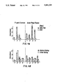
FIG. 7. Detection of Ag-7H8 in the plasma samples of various strains of mice infected with 17XNL strain of P. voelii. Groups of nine mice each were infected with 106 17XNL P. voelii parasites. Parasitemias (the left axis and ascending curves) are the mean for six to nine animals. Blood samples, collected from three mice on the days indicated, were pooled and assayed in the two-sited assay. Results (the right axis and bars) are expressed as means ±1 standard deviation for triplicate samples. Results for all C57B1/6 (B6) congenics were virtually identical, so only one is shown.
FIG. 8. Indirect Immunofluorescence pattern produced by normal mouse serum (negative control), immune serum on rings, trophozoites and schizonts, and MAb 7H8 on P. voelii and P. falciparum.
FIG. 9. Western blot analysis of antigen 7H8 in P. berghei (Pb), P. voelli (Py), and P. falciparum (Pf).
FIG. 10. Schematic diagram showing production of a portion of antigen Pf93 in the lambda gt11/E. coli Y1090 system.
FIG. 11. Flow diagram showing steps of production and release of Pf93 in P. falciparum.
FIG. 12. IFA analysis of course of Pf93 production after RBC infection.
FIG. 13. Schematic diagram showing two-sited assay using MAb 7H8.
FIG. 14. Time course of antigen release from infected RBC's determined by two-sited assay using MAb 7H8.
FIG. 15. Antigen release following reinvasion of RBC's determined by two-sited assay using MAb 7H8.
FIG. 16. Detection by two-sited assay using MAb 7H8 of antigen Pf93 in sera from individuals uninfected and acutely infected with P. falciparum.
FIG. 17. In vitro inhibition of merozoite invasion in human erythrocytes by MAb 7H8.
FIG. 18. Graph showing reduced parasitemia in 17XNL P. voelli malaria-challenged BALB/c mice immunized with anti-idiotypic antibody to MAb 7H8.
FIG. 19. ELISA of first fusion culture supernatants for anti-idiotypic antibodies to MAb 7H8. The asterisk denotes ELISA-positive culture containing antibody against MAb 7H8. (Horizontal line=25% of maximum specific binding.)
FIG. 20. ELISA of second fusion culture supernatants for anti-idiotypic antibodies to MAb 7H8. The asterisk denotes ELISA-positive culture containing antibody against MAb 7H8. (Horizontal line=25% of maximum specific binding.)
FIG. 21. Visualization of P. voelli 17XNL parasites treated with anti anti-idiotypic antibodies in sera of mice immunized with anti-idiotypic antibodies to MAb 7H8. (A) Fluorescence microscopy. (B) Nomarski differential interference contrast microscopy.
DETAILED DESCRIPTION OF THE PREFERRED EMBODIMENTS
The present invention relates to a monoclonal antibody (MAb 7H8) which identifies a pan-malarial antigen (Ag-7H8). This MAb is produced by a continuous hybridoma cell line (cell line 7H8) generated by infecting BALB/c mice with 104 17XL P. voelii parasites. This monoclonal antibody is well suited for immunological detection of species of the genus Plasmodium, including P. voelii, P. berghei, P. falciparum, P. vinckei, P. chabaudi, P. knowlesi, P. gallinaceum, P. vivax, P. ovale, and P. malariae. MAb 7H8 is secreted by a continuous hybridoma cell line, deposited at the American Type Culture Collection, 12301 Parklawn Drive, Rockville, Maryland 20852, on Dec. 5, 1986, having accession number HB 9287. Thus, in one embodiment, the present invention provides for a continuous hybridoma cell line characterized in that it is capable of producing a monoclonal antibody having the specificity of the monoclonal antibody produced by hybridoma cell line 7H8. In another embodiment, the present invention provides for a continuous hybridoma cell line capable of secreting a monoclonal antibody reactive with an antigen (Ag-7H8), said antigen characterized in that:
(1) the major form of the antigen has a molecular weight of about 120,000, with minor components of 220,000, 65,000 to 75,000, and 45,000 in P. veolii;
(2) it remains antigenic after boiling for five minutes; and
(3) said antigen can be bound by monoclonal antibody 7H8.
In yet another embodiment, the present invention provides for a monoclonal antibody having the specificity of MAb 7H8, said MAb 7H8 characterized in that it is produced by the hybridoma cell line 7H8.
The inventor has also discovered that MAb 7H8 identifies an antigen that is unique to one particular species of Plasmodium, namely P. falciparum. This antigen, designated Pf93, may be isolated in substantially pure form, employing MAb 7H8 of the present invention. Thus, an embodiment of the present invention provides for substantially pure antigen Pf93, said antigen characterized in that it is recognized by and binds to MAb 7H8. In another embodiment, the present invention provides for a monoclonal antibody capable of binding to the Pf93 antigen, said monoclonal antibody characterized in that it has the specificity of monoclonal antibody 7H8. In yet another embodiment, the present invention provides for a substantially purified P. falciparum antigen obtainable by:
(a) solubilizing P. falciparum cells containing said antigen;
(b) contacting the thus solubilized P. falciparum of step (a) with a monoclonal antibody having the specificity of MAb 7H8, thereby forming a complex of said antigen and said monoclonal antibody;
(c) substantially isolating said complex; and
(d) separating said antigen from said monoclonal antibody.
The substantially pure antigen Pf.93 of the present invention may in turn be used to detect or measure antibody to plasmodial infection by P. falciparum in a sample, such as serum. Thus, one embodiment of the present invention comprises a method of detecting the presence or amount of antibody to plasmodium-associated antigen in a sample, comprising contacting said sample containing said antibody to plasmodium-associated antigen with detectably-labeled Pf93, and detecting said label. In addition to antigen Pf93 itself, one may also use immunoreactive fractions and immunoreactive analogues of Pf93. By the term "immunoreactive reactive fractions(s)" is intended a portion of the Pf93 antigen which demonstrates the equivalent immune response to the 7H8 antibody. By the term "immunoreactive analogue" is intended a protein which differs from the Pf93 protein by one or more amino acids but which demonstrates the equivalent immune response to antibody 7H8.
MAb 7H8 is well suited for use in standard immunodiagnostic assays well-known in the art, including such immunometric or "sandwich" assays as the forward sandwich, reverse sandwich, and simultaneous sandwich assays In this regard, the monoclonal antibody of the present invention may be used in combination with one or more other monoclonal antibodies specific for the Plasmodium genus or for particular Plasmodium species. The present inventor has discovered that the antigen 7H8 has at least two similar epitopes per molecule. As a result, a two-sited assay developed by the present inventor is a preferred form of immunoassay according to the present invention. Thus, in one embodiment, the present invention provides a method of determining the presence or amount of plasmodium-associated antigen in a sample, comprising:
(a) binding MAb 7H8 to an insoluble phase;
(b) adding a sample suspected of containing soluble plasmodium-associated antigen;
(c) adding detectably labeled MAb 7H8 to the soluble phase;
(d) removing unbound detectably labeled MAb 7H8; and
(e) detecting said label.
In yet another embodiment, the present invention provides a method of determining the presence or amount of plasmodium-associated antigen in a sample comprising:
(a) binding partially purified MAb 7H8 to microtiter wells;
(b) adding either parasite extract or plasma samples containing soluble antigens;
(c) adding alkaline-phosphatase-labeled MAb 7H8; and
(d) adding a substrate and recording the subsequent amount of color development.
Although the inventor's present data suggest that there may be no therapeutic utility, the antibody and substantially purified antigens of the present invention also may be useful for the therapeutic treatment of plasmodial infections. Thus, one embodiment of the present invention provides a method of suppressing a plasmodial infection in an animal which comprises administering to said animal a therapeutically effective amount of a monoclonal antibody having the specificity of MAb 7H8. In yet another embodiment, the present invention provides a pharmaceutical composition comprising plasmodial infection-suppressing amounts of MAb 7H8, together with a pharmaceutically inert carrier.
Those of skill will appreciate that preventing plasmodial infection is greatly to be preferred where possible. Using the monoclonal antibody of the present invention, it may be possible to immunize an animal, such as a human, against plasmodial infection. Thus, one embodiment of the present invention provides a method of immunizing an animal against plasmodial infection which comprises administering to said animal an effective amount of a monoclonal antibody having the specificity of MAb 7H8. It will also be appreciated that anti-idiotypic and anti anti-idiotypic antibodies to MAb 7H8 may be generated, which will be useful for vaccines, passive immunization, and the like. Thus, another embodiment of the present invention comprises an anti-idiotypic antibody to MAb 7H8. Yet another embodiment of the present invention comprises an anti anti-idiotypic antibody to MAb 7H8. A further embodiment of the present invention comprises a method of actively immunizing an animal against plasmodial infection comprising administering to said animal effective amounts of an anti-idiotypic antibody to MAb 7H8 or passively immunizing an animal by administering effective amounts of an anti anti-idiotypic antibody to MAb 7H8.
In the following description, reference will be made to various methodologies well known to those skilled in the art of immunology. Applications and other materials setting forth such well-known methodologies will be referred to in the course of this description, and are incorporated herein by reference in their entirety as set forth herein in full. Standard reference works setting forth the general principles of immunology include Klein, J. Immunology: The Science of Self-Nonself Discrimination, John Wiley & Sons, Publisher, New York (1982); Kennett, R., et al., eds., Monoclonal Antibodies, Hybridoma: A New Dimension in Biological Analyses, Plenum Press, Publisher, New York (1980); Campbell, A., "Monoclonal Antibody Technology", in, Burdon, R., et al., eds., Laboratory Techniques in Biochemistry and Molecular Biology, Volume 13, Elsevier, Publisher, Amsterdam (1984).
By "detecting" it is intended to include determining the presence or absence of a substance or quantifying the amount of a substance. The term thus refers to the use of the materials, compositions, and methods of the present invention for qualitative and quantitative determinations.
By "plasmodial infection" is meant any infection of an animal, including a human, caused by any species of the genus Plasmodium. A preferred species for the purposes of the present invention is P. falciparum.
By "plasmodium-associated antigen" is meant any antigen associated with a species of the genus Plasmodium, which antigen is expressed during the course of infection in its animal host. Such antigens may be associated with the surface of the parasite, or may be released by the parasite into the host RBC and then into serum. The latter are preferred for the purposes of the present invention. Particularly preferred is the so-called 7H8 family of conserved antigens. These antigens are expressed by many different Plasmodium species, and are recognized by MAb 7H8. They are thus referred to as the "pan-malarial" antigens. Even more preferred is the Pf93 antigen, which is unique to P. falciparum.
The procedures for the production of the monoclonal antibody of the present invention are described in Taylor, D.W., et al., Infection Immun. 32(a):563-570 (1981), incorporated herein by reference. Briefly, BALB/c mice were infected with 17XL P. voelii parasites. Mice were killed when parasitemias reached about 54%, and a suspension of spleen cells was prepared and fused with the P3-X63-NS/1 cell line. Hybrids were selected in a hypoxanthine-aminopterinethymidine medium and screened by indirect immunofluorescence (IIF) with acetone-fixed smears of P. voelii and by radioimmunoassay as described by Taylor, D.W., et al., Exp. Parasitol. 53:362-370 (1981), with extracts of 17XL and 17XNL parasites. The hybrid cell line (7H8) used in this study was cloned twice by limiting dilution. Ascites was produced in pristane-primed mice. The isotype of the hybrid was determined by polyethylene glycol Ouchterlony analysis, immunoelectrophoresis, and an isotype-specific radioimmunoassay as described by Taylor, D.W., et al., Exp. Parasitol. 53:362-370 (1981).
The isolation of other hybridomas secreting monoclonal antibodies of the same specificity as those described herein can be accomplished by the technique of anti-idiotypic screening. Potocnjak, et al., Science 215:1637 (1982). Briefly, an anti-idiotypic antibody is an antibody which recognizes unique determinants present on the antibody produced by the clone of interest. The anti-idiotypic antibody is prepared by immunizing an animal of the same strain used as the source of the monoclonal antibody with the monoclonal antibody of interest. The immunized animal will recognize and respond to the idiotypic determinants of the immunizing antibody by producing antibody to these idiotypic determinants (anti-idiotypic antibody). By using the anti-idiotypic antibody of the second animal, which is specific for the monoclonal antibodies produced by a single clone, it is then possible to identify other clones used for immunization. Idiotypic identity between the product of two clones demonstrates that the two clones are identical with respect to their recognition of the same epitopic determinants. The anti-idiotypic antibody may also be used as an "immunogen" to induce an immune response in yet another animal, producing a so-called anti anti-idiotypic antibody which will be epitopically identical to the original MAb. Thus, by using antibodies to the epitopic determinants of a monoclonal antibody, it is possible to identify other clones expressing antibodies of identical epitopic specificity. In antibodies, idiotypic determinants are present in the hypervariable region which binds to a given epitope
Accordingly, the monoclonal antibody of the present invention may be used to induce anti-idiotypic Abs in BALB/c mice. Spleen cells from these animals are used to produce anti-idiotypic hybridoma cell lines. Monoclonal anti-idiotypic Abs coupled to KLH are used as "immunogen" to immunize BALB/c mice. Sera from these mice will contain anti anti-idiotypic Abs that have the binding properties of the original Ab specific for the shared epitope. The anti-idiotypic MAbs thus have idiotopes structurally similar to the epitope being evaluated.
For replication, the hybrid cells may be cultivated both in vitro and in vivo. High in vivo production makes this the presently preferred method of culture. Briefly, cells from the individual hybrid strains are injected intraperitoneally into pristane-primed BALB/C mice to produce ascites fluid containing high concentrations of the desired monoclonal antibodies. Monoclonal antibodies of isotype IgM may be purified from cultured supernatants using column chromatography methods well known to those of skill in the art.
The monoclonal antibody of the present invention is particularly suited for use in immunoassays wherein it may be utilized in liquid phase or bound to a solid phase carrier. In addition, the monoclonal antibody in these immunoassays can be detectably labeled in various ways.
The term "antibody" (Ab) as used in this invention is meant to include intact molecules as well as fragments thereof, such as, for example, Fab and F(ab')2 ' which are capable of binding antigen. Fab and F(ab')2 fragments lack the Fc fragment of antibody, clear more rapidly from the circulation, and may have less nonspecific tissue binding than intact antibody. Wahl et al., J. Nuclear Med. 24(4):316-325 (1983). It will be appreciated that Fab, F(ab')2 and other fragments of the monoclonal antibody of the present invention may be used as well as the intact antibody for the detection and treatment of plasmodial antigens according to the methods of the present invention.
There are many different labels and methods of labeling known of those of ordinary skill in the art. Examples of the types of labels which can be used in the present invention include, but are not limited to, enzymes, radioisotopes, fluorescent compounds, chemiluminescent compounds, bioluminescent compounds and metal chelates. Those of ordinary skill in the art will know of other suitable labels for binding to the monoclonal antibody, or will be able to ascertain the same by the use of routine experimentation. Furthermore, the binding of these labels to the monoclonal antibody can be accomplished using standard techniques commonly known to those of ordinary skill in the art.
One of the ways in which a monoclonal antibody of the present invention can be detectably labeled is by linking the monoclonal antibody to an enzyme. This enzyme, in turn, when later exposed to its substrate, will react with the substrate in such a manner as to produce a chemical moiety which can be detected as, for example, by spectrophotometric or fluorometric means. Examples of enzymes which can be used to detectably label the monoclonal antibodies of the present invention include malate dehydrogenase, staphyloccocal nuclease, delta-V-steroid isomerase, yeast alcohol dehydrogenase, alpha-glycerophosphate dehydrogenase, triose phosphate isomerase, horseradish peroxidase, alkaline phosphatase, asparaginase, glucose oxidase, beta-galactosidase, ribonuclease, urease, catalase, glucose-VI-phosphate dehydrogenase, glucoamylase and acetylcholine esterase.
The presence of the detectably labeled monoclonal antibody of the present invention can also be detected by labeling the monoclonal antibody with a radioactive isotope which can then be determined by such means as the use of a gamma counter or a scintillation counter. Isotopes which are particularly useful for the purpose of the present invention are 3 H, 125 I, 32 P, 35 S, 14 C, 51 Cr, 36 Cl, 57 Co, 58 Co, 59 Fe and 75 Se.
It is also possible to detect the binding of the detectably labeled monoclonal antibody of the present invention by labeling the monoclonal antibody with a fluorescent compound. When the fluorescently labeled monoclonal antibody is exposed to light of the proper wave length, its presence can then be detected due to the fluorescence of the dye. Among the most commonly used fluorescent labelling compounds are fluoroscein isothiocyanate, rhodamine, phycoerytherin, phycocyanin, allophycocyanin, o-phthaldehyde and fluorescamine.
The monoclonal antibody of the invention also can be detectably labeled using fluorescent emitting metals such as 152 Eu, or others of the lanthanide series. These metals can be attached to the antibody molecule using such metal chelating groups as diethylenetriaminepentaacetic acid (DTPA) or ethylenediaminetetraacetate acid (EDTA).
The monoclonal antibody of the present invention also can be detectably labeled by coupling it to a chemiluminescent compound. The presence of the chemiluminescent-tagged monoclonal antibody is then determined by detecting the presence of luminescence that arises during the course of the chemical reaction. Examples of particularly useful chemiluminescent labeling compounds are luminol, isoluminol, theromatic acridinium ester, imidazole, acridinium salt and oxalate ester.
Likewise, a bioluminescent compound may be used to label the monoclonal antibody of the present invention. Bioluminescence is a type of chemiluminescence found in biological systems in which a catalytic protein increases the efficiency of the chemiluminescent reaction. The presence of a bioluminescent monoclonal antibody is determined by detecting the presence of luminescence. Important bioluminescent compounds for purposes of labeling are luciferin, luciferase and aequorin.
For the purposes of the present invention, plasmodial-associated antigen which is detected by the monoclonal antibody of the invention may be present in biological fluids and tissues. Any sample containing the detectable yet unknown amount of plasmodial-associated antigen can be used. Normally, the sample is a liquid, such as, for example, urine, saliva, cerebrospinal fluid, blood, serum and the like, or a solid or semi-solid, such as, for example, tissue, feces and the like.
Another technique which may also result in greater sensitivity when used in conjunction with the present invention consists of coupling the monoclonal antibody of the present invention to low molecular weight haptens. The haptens can then be specifically detected by means of a second reaction. For example, it is common to use such haptens as biotin (reacting with avidin) or dinitrophenyl, pyridoxal and fluorescamine (reacting with specific antihapten antibodies) in this manner.
The term "epitope" as used in this invention is meant to include any determinant responsible for specific interaction with an antibody molecule. Epitopic determinants usually consist of chemically active surface groupings of molecules such as amino acids or sugar side chains and have specific three-dimensional structural characteristics, as well as specific charge characteristics.
For in vivo diagnosis, radionucleotides may be bound to the monoclonal antibodies of the present invention either directly or by using an intermediary functional group. An
intermediary group which is often used to bind radioisotopes which exist as metallic cations to antibodies is diethylenetriaminepentaacetic acid (DTPA). Typical examples of metallic cations which are bound in this manner are: 99m Tc, 123 I, 111 IN, 131 I, 97 Ru, 67 Cu, 67 Ga and 68 Ga The monoclonal antibodies of the invention can also be labeled with non-radioactive isotopes for purposes of in vivo diagnosis. Elements which are particularly useful in this manner are 157 Gd, 55 Mn, 162 52 Cr and 56 Fe.
The monoclonal antibody of the present invention also may be used for immunotherapy in animals, including humans, having a malarial infection which expresses plasmodial-associated antigen with epitopes reactive with the monoclonal antibody of the present invention. When used for immunotherapy, the monoclonal antibody of the present invention may be unlabeled or labeled with a therapeutic agent. Examples of therapeutic agents which can be coupled to the monoclonal antibodies of the invention for immunotherapy are drugs, radioisotopes, lectins and toxins.
Lectins are proteins, usually isolated from plant material, which bind to specific sugar moieties. Many lectins are also able to agglutinate cells and stimulate lymphocytes. Ricin is a toxic lectin which has been used immunotherapeutically. This use is accomplished by binding the alpha-peptide chain of ricin, which is responsible for toxicity, to the antibody molecule to enable site-specific delivery of the toxic defect.
Toxins are poisonous substances produced by plants, animals or microorganisms that, in sufficient dose, are often lethal. Diphtheria toxin, for example, is a protein produced by Corynebacterium diphtheria. This toxin consists of an alpha and a beta subunit which under proper conditions can be separated. The toxic alpha component can be bound to antibody and used for a site-specific delivery.
Examples of radioisotopes which can be bound to the monoclonal antibodies of the present invention for use in immunotherapy are: 125 Um, 131 I, 90 Y, 67 Cu, 217 Bi, 211 At, 212 Pb, 47 Sc and 109 Pd.
It is also possible to utilize liposomes having the monoclonal antibody of the present invention in their membrane to specifically deliver the liposomes to the target area. These liposomes can be produced so that they contain, in addition to the monoclonal antibody, such immunotherapy agents as drugs, radioisotopes, lectins and toxins, which would be released at the target site.
The dose ranges for the administration of the monoclonal antibody of the present invention are those large enough to produce the desired effect, whereby the malarial symptoms are ameliorated. The doses should not be so large as to cause adverse side effects, such as unwanted cross reactions, anaphylactic reactions, and the like. Generally, the dosage will vary with the age, condition, sex and extent of the disease in the patient. Counterindications, if any, immune tolerance and other variables will also affect the proper dosage. The monoclonal antibody can be administered parenterally by injection or by gradual profusion over time. The monoclonal antibody of the present invention also can be administered intravenously, intraparenterally, intramuscularly or subcutaneously.
Preparations for parenteral administration include sterile or aqueous or non-aqueous solutions, suspensions and emulsions. Examples of non-aqueous solvents are propylene glycol, polyethylene glycol, vegetable oils such as olive oil, and injectable organic esters such as ethyloleate Aqueous carriers include water, alcoholic/aqueous solutions, emulsions or suspensions, including saline and buffered media. Parenteral vehicles include sodium chloride solution, Ringer's dextrose, dextrose and sodium chloride, lactated Ringer's, or fixed oils. Intravenous vehicles include fluid and nutrient replenishers, electrolyte replenishers, such as those based on Ringer's dextrose, and the like. Preservatives and other additives may also be present, such as, for example, antimicrobials, anti-oxidants, chelating agents, inert gases and the like. See, generally, Reminqton's Pharmaceutical Science, 16th ed., Mac Eds. (1980).
The present invention also relates to a method of preparing a medicament or a pharmaceutical composition comprising the components of the invention, the medicament being used for therapy of animal, including human, malaria-expressing, or fractions or analogues thereof, antigen reactive with the monoclonal antibody of the present invention.
The monoclonal antibody and substantially purified antigen of the present invention are ideally suited for the preparation of a kit. Such a kit may comprise a carrier means being compartmentalized to receive in close confinement therewith one or more container means such as vials, tubes and the like, each of said container means comprising the separate elements of the immunoassay to be used.
The types of immunoassays which can be incorporated in kit form are many. Typical examples of some of the immunoassays which can utilize the antibodies of the invention are radioimmunoassays (RIA) and immunometric, or sandwich, immunoassays.
By the term "immunometric assay" or "sandwich immunoassay", it is meant to include simultaneous sandwich, forward sandwich and reverse sandwich immunoassays. These terms are well understood by those skilled in the art. Those of skill will also appreciate that the monoclonal antibody of the present invention will be useful in other variations and forms of immunoassays which are presently known or which may be developed in the future. These are intended to be included within the scope of the present invention
In a forward sandwich immunoassay, a sample is first incubated with a solid phase immunoabsorbent containing monoclonal antibody(ies) against the antigen Incubation is continued for a period of time sufficient to allow the antigen in the sample to bind to the immobilized antibody in the solid phase. After the first incubation, the solid phase immunoabsorbent is separated from the incubation mixture and washed to remove excess antigen and other interfering substances, such as non-specific binding proteins, which also may be present in the sample. Solid phase immunoabsorbent containing antigen bound to the immobilized antibody is subsequently incubated for a second time with soluble labeled antibody or antibodies. After the second incubation, another wash is performed to remove unbound labeled antibody(ies) from the solid phase immunoabsorbent and removing non-specifically bound labeled antibody(ies). Labeled antibody(ies) bound to the solid phase immunoabsorbent is then detected and the amount of labeled antibody detected serves as a direct measure of the amount of antigen present in the original sample. Alternatively, labeled antibody which is not associated with the immunoabsorbent complex can also be detected, in which case the measure is in inverse proportion to the amount of antigen present in the sample. Forward sandwich assays are described, for example, in U.S. Pat. Nos. 3,867,517; 4,012,294 and 4,376,110.
In carrying out forward immunometric assays, the process comprises, in more detail:
(a) first forming a mixture of the sample with the solid phase bound antibody(ies) and incubating the mixture for a time and under conditions sufficient to allow antigen in the sample to bind to the solid phase bound antibody(ies).
(b) adding to the mixture after said incubation of step
(a) the detectably labeled antibody or antibodies and incubating the new resulting mixture for a time and under conditions sufficient to allow the labeled antibody to bind to the antigen-antibody complex on the solid phase immunoabsorbent;
(c) separating the solid phase immunoabsorbent from the mixture after the incubation in step (b); and
(d) detecting either the labeled antibody or antibodies bound to the antigen-antibody complex on the solid phase immunoabsorbent or detecting the antibody not associated therewith.
In a reverse sandwich assay, the sample is initially incubated with labeled antibody(ies), after which the solid phase immunoabsorbent containing multiple immobilized antibodies is added thereto, and a second incubation is carried out. The initial washing step of a forward sandwich assay is not required, although a wash is performed after the second incubation. Reverse sandwich assays have been described, for example, in U.S. Pat. Nos. 4,098,876 and 4,376,110.
In carrying out reverse immunometric assays, the process comprises, in more detail;
(a) first forming a mixture of the sample with the soluble detectably labeled antibody for a time and under conditions sufficient to allow antigen in the sample to bind to the labeled antibody;
(b) adding to the mixture after the incubation of step (a) the solid phase bound antibodies and incubating the new resulting mixture for a time and under conditions sufficient to allow antigen bound to the labeled antibody to bind to the solid phase anti
bodies;
(c) separating the solid phase immunoabsorbent from the incubating mixture after the incubation in step (b); and
(d) detecting either the labeled antibody bound to the solid phase immunoabsorbent or detecting the labeled antibody not associated therewith.
In a simultaneous sandwich assay, the sample, the immunoabsorbent having multiple immobilized antibodies thereon and labeled soluble antibody or antibodies are incubated simultaneously in one incubation step. The simultaneous assay requires only a single incubation and has a lack of washing steps. The use of a simultaneous assay is by far the preferred one. This type of assay brings about ease of handling, homogeneity reproducibility, linearity of the assays and high precision. The sample containing antigen, solid phase immunoabsorbent with immobilized antibodies and labeled soluble antibody or antibodies is incubated under conditions and for a period of time sufficient to allow antigen to bind to the immobilized antibodies and to the soluble antibody(ies). In general, it is desirable to provide incubation conditions sufficient to bind as much antigen as possible, since this maximizes the binding of labeled antibody to the solid phase, thereby increasing the signal. Typical conditions of time and temperature are two hours at 45° C., or twelve hours at 37° C.
Antigen typically binds to labeled antibody more rapidly than to immobilized antibody, since the former is in solution whereas the latter is bound to the solid phase support. Because of this, labeled antibody may be employed in a lower concentration than immobilized antibody, and it is also preferable to employ a high specific activity for labeled antibody. For example, labeled antibody might be employed at a concentration of about 1-50 ng per assay, whereas immobilized antibody might have a concentration of 10-500 ng per assay per antibody. The labeled antibody might have a specific activity with, for instance, one radioiodine per molecule, or as high as two or more radioiodines per molecule of antibody.
Of course, the specific concentrations of labeled and immobilized antibodies, the temperature and time of incubation as well as other assay conditions can be varied, depending on various factors including the concentration of antigen in the sample, the nature of the sample and the like. Those skilled in the art will be able to determine operative and optimal assay conditions for each determination by employing routine experimentation.
After the single incubation period, the solid phase immunoabsorbent is removed from the incubation mixture. This can be accomplished by any of the known separation techniques, such as sedimentation and centrifugation. A washing step is not required prior to detection of bound labeled antibody. Detection can be performed by a scintillation counter, for example, if the label is a radioactive gamma-emitter, or by a fluorometer, for example, if the label is a fluorescent material. In the case of an enzyme label, the detection can be done by colorimetric methods employing a substrate for the enzyme.
In carrying out the simultaneous immunometric assay on a sample containing a multivalent antigen, the process comprises, in more detail:
(a) simultaneously forming a mixture comprising the sample, together with the solid phase bound antibody and the soluble labeled antibody or antibodies;
(b) incubating the mixture formed in step (a) for a time and under conditions sufficient to allow antigen in the sample to bind to both immobilized and labeled antibodies;
(c) separating the solid phase immunoabsorbent from the incubation mixture after the incubation; and
(d) detecting either labeled antibody bound to the solid phase immunoabsorbent or detecting labeled antibody not associated therewith.
Other such steps as washing, stirring, shaking filtering and the like may of course be added to the assays, as is the custom or necessity for any particular situation.
In the preferred mode for preforming the assays it is important that certain "blockers" be present in the incubation medium (usually added with the labeled soluble antibody). The "blockers" are added to assure that non-specific proteins, protease, or human antibodies to mouse immunoglobulins present in the experimental sample do not cross-link or destroy the monoclonal antibodies on the solid phase support, or the radiolabeled indicator antibody, to yield false positive or false negative results. The selection of "blockers" therefore adds substantially to the specificity of the assays described in the present invention.
It has been found that a number of nonrelevant (i.e., nonspecific) monoclonal antibodies of the same class or subclass (isotype) as those used in the assays (e.g. IgG1, IgG2a, IgM, etc.) can be used as "blockers". The concentration of the "blockers" (normally 1-100,ug/ul) is important, in order to maintain the proper sensitivity yet inhibit any unwanted interference by mutually occurring cross reactive proteins in human serum. In addition, the buffer system containing the "blockers" needs to be optimized. Preferred buffers are those based on weak organic acids, such as imidazole, HEPPS, MOPS, TES, ADA, ACES, HEPES, PIPES, TRIS, and the like, at physiological pH ranges. Somewhat less preferred buffers are inorganic buffers such as phosphate, borate or carbonate. Finally, known protease inhibitors should be added (normally at 0.01-10 microgs/ml) to the buffer which contains the "blockers".
There are many solid phase immunoabsorbents which have been employed and which can be used in the present invention. Well known immunoabsorbents include glass, polystyrene, polypropylene, dextran, nylon and other materials; tubes, beads, and microtiter plates formed from or coated with such materials, and the like. The immobilized antibodies can be either covalently or physically bound to the solid phase immunoabsorbent, by techniques such as covalent bonding via an amide or ester linkage, or by absorption. Those skilled in the art will know many other suitable solid phase immunoabsorbents and methods for immobilizing antibodies thereon, or will be able to ascertain such, using no more than routine experimentation.
The materials and methods used in carrying out the present invention may be more fully understood by reference to the following examples, which examples are not intended in any manner to limit the scope of the present invention.
One aspect of the present invention involves the development of pan-malarial vaccines. There are various methods for preparing vaccines against viruses and bacteria. The basic preferred requirements for any vaccine and for a method for the preparation of a vaccine are that (1) the resulting vaccine contain the necessary antigenic determinants to induce formation of antibodies in the host; (2) the vaccine possess high immunogenic potential; (3) the resulting vaccine be safe enough to be administered without any danger of clinical infection, either for the recipient or any contact of the recipient, and therefore, the risk associated with vaccination be minimized, if not totally eliminated; (4) the resulting vaccine be devoid of any toxic side-effects, for example, fever from endotoxin present in killed or extracted cells; (5) the resulting vaccine be suitable for administration by an effective route, for example, oral, intranasal, topical or parenteral; (6) the resulting vaccine mimic closely the circumstances of natural infection; (7) the resulting vaccine be stable under conditions of long-term storage, and that said long-term storage be at room temperature; and (8) the resulting vaccine be compatible with the usual inert vaccine carriers. Those conditions can be achieved by the vaccines of the present invention.
In one embodiment of the present invention, vaccines prepared from PfΣantigen, its immunogenic fraction or analogue, or anti-idiotypic antibody to 7H8 antibody, comprise the antigenic component of the vaccine. It may be necessary or preferable to covalently link the antigenic component to an immunogenic carrier, i.e., bovine serum albumin or keyhole limpet hemocyanin. The vaccines of the present invention may be administered to any mammal susceptible to plasmodial infection. Human and non-human mammals may benefit as hosts.
Administration may be by parenteral, oral, intranasal, intravenous, intramuscular, subcutaneous, or any other suitable means. The dosage administered may be dependent upon, the age, health, weight, kind of concurrent treatment, if any, and nature of the plasmodial organism. The vaccine may be employed in dosage forms such as capsules, liquid solutions, suspensions, or elixirs, for oral administration, or sterile liquid formulations such as solutions or suspensions. An inert, immunologically acceptable carrier is preferably used, such as saline or phosphate-buffered saline.
EXAMPLE I MATERIALS AND METHODS Strains of Mice and Parasites
In the majority of the studies, 6- to 8-week-old BALB/c mice (Cumberland View Farms, Clinton, Tenn.) were injected intraperitoneally (i.p.) with specified numbers of either the 7XL (lethal) or 17XNL (nonlethal) strain of P. voelii. Parasitemias were monitored every other day from Giemsa-stained tail blood smears. In studies comparing the kinetics of parasitemia and antigenemia, mice of the following strains were injected i.p. with 106 17XNL P. voelii parasites: AKR/J (Jackson Laboratory, Bar Harbor, Me.), C57BL/6Boy and C57BL/6(H-2k) (NIAID breeding contact; Bioqual Inc., Rockville, MD). Parasites used in indirect immunofluorescence (IIF) studies included Plasmodium berghei (NYU/2) and P. falciparum (Malayan Camp strain) from Aotus monkeys.
Production of MAbs
Details of the procedures for the production of anti-P. voelii MAbs are described in, Taylor, D.W. et al., Infect. Immun. 32:563-570 (1981), incorporated herein by reference. Briefly, the hybridoma of the present invention was produced by infecting BALB/c mice with 104 17XL P. voelli parasites. When the parasitemias reached 54% 9 days later, the mice were killed, and a suspension of spleen cells was prepared and fused with the P3-X63-NS/1 cell line. Hybrids were selected in a hypoxanthine-aminopterin-thymidine medium and screened by IIF with acetone-fixed smears of P. voelli, Voller, A., Bull. W.H.O. 30:343-354, and by radioimmunoassay as described in, Taylor, D.W. et al., Exp. Parasitol 53:362-370 (hereinafter Taylor (I)), with extracts of 17XL and 17XNL parasites. The hybrid cell line (7H8) of the present invention was cloned twice by limiting dilution. Ascites was produced in pristane-primed mice. The isotype of the hybrid was determined by polyethylene glycol Ouchterlony analysis, immunoelectrophoresis, and an isotype-specific radioimmunoassay as previously described. Taylor,(I).
Molecular Weight of Ag-7H8
P. voelii parasites of the 17XL strain were cultured in vitro for 14 h in Selectamine (GIBCO Laboratories, Grand Island, N.Y.) in the presence of 1 uCi of [35 S] methionine as described in Langhorne, J., et al., Cell. Immunol. 89:452-461. Cells were harvested, washed three times with saline, and solubilized with 0.5% Nonidet P-40 in NET buffer (0.15 M NaCl, 2 mM EDTA, 0.5 M Tris: pH 7.4). Aliquots of labeled parasite proteins were adsorbed with protein A-Sepharose (Pharmacia Fine Chemicals, Piscataway, N.J.) and then incubated for 30 min with 5 ul of (i) normal mouse serum; (ii) immune mouse serum; (iii) ascites from the MAb 8B11 known to react with a protein of 230,000 Mr in P. voelli; and (iv) MAb 7H8. Next, 50 ul of a 1:20 dilution of rabbit anti-mouse mu serum was added and incubated for 30 min at room temperature. Finally, 50 ul of a 50% slurry of protein A-Sepharose in NET buffer was added and the mixture was incubated for 45 min. Antigen-antibody-protein-A-Sepharose complexes were washed extensively, solubilized in sodium dodecyl sulfate (SDS)-sample buffer (nonreducing), and electrophoresed on a 10% slab SDS-polyacryl-amine gel (SDS-PAG) by the method of Laemmli. Laemmli, U.K., Nature 227:680-682 by the method of Laemmli. (1970). Gels were treated with En3 Hance (New England Nuclear Corp., Boston, Mass.), dried, and processed for autoradiography with X-Omat R film (Eastman Kodak Co., Rochester, N.Y.).
Molecular weight determinations were also made by Western blot analysis as described in Howard, R.J., J. Cell Biol. (in press). Extracts of 17XL P. voelli, prepared by freeze-thawing Percoll-enriched, P. voelli-infected erythrocytes, were incubated for 10 min at 37° C. in 5% SDS-5% 2-mercaptoethanol and then electrophoresed on a 5 to 15% SDS-PAG according to Laemmli. Laemmli, U.K., Nature 227:680-682 (1970). Proteins were transblotted onto nitrocellulose paper, Towbin, H., et al., Proc. Natl. Acad. Sci. USA 76:43-50-4354 (1979), overnight in a Hoefer apparatus (Hoefer Scientific Instruments, San Francisco, Calif.) at 30 V with Tris-glycine buffer plus 20% methanol. Nitrocellulose strips were then incubated successively with 2 ml of 0.3% Tween 20 plus 0.3% bovine serum albumin (BSA) in NET buffer, a 1:100 dilution of MAbs, a 1:1,000 dilution of rabbit anti-mouse immunoglobulin M (IgM), and 125 I-protein A (30,000 cpm/ml; ICN Pharmaceuticals, Inc., Irvine, Calif.). Strips were dried on gel bond and autoradiographed as described above. High- and low-molecular weight standards (Bio-Rad Laboratories, Richmond, Calif.), were used throughout.
Antigen-Detection Assay
A two-sited assay was developed for detecting Ag-7H8 in the sera of infected mice. This assay consists of four major steps: (i) binding of partially purified MAb 7H8 to microtiter wells, (ii) adding either parasite extract or plasma samples containing soluble antigens, (iii) adding alkaline-phosphatase-labeled MAb 7H8, and finally (iv) adding a substrate and recording the subsequent amount of color development. Initially, MAb 7H8 was partially purified by a 50% ammonium sulfate precipitation. Then, 1 mg was coupled to 1,000 U of alkaline phosphate (Sigma Chemical Co., St. Louis, Mo.) by incubation with 0.25% glutaraldehyde for 2 h at room temperature, followed by extensive dialysis against phosphate-buffered saline (PBS), pH 7.4. In the two-sited assay, 100 ul of partially purified MAb 7H8 in PBS, at 100 ug/ml unless otherwise specified, was applied to the wells of flat-bottom polystyrene microtiter plates (Immunolon I; Dynatech Industries, Inc., McLean, Va.), and control wells were treated with 3% BSA (radioimmunoassay grade) in PBS. Plates were incubated overnight at 4° C. Wells were washed, treated with 200 ul of 3% BSA in PBS for 2 h at 37° C., and washed five times with PBS. Then, 50 ul of P. voelii extract, plasma samples diluted in 3% BSA in PBS, or PBS (used as a negative control for blanking the microplate reader) was added to triplicate wells and incubated overnight. After being washed five times with PBS, 100 ul of alkaline-phosphate-labeled 7H8 MAb (at a 1:500 dilution unless otherwise stated) was added. Plates were incubated for 2 h at 37° C. and washed three times with PBS, and 200 ul of 1 mg of p-nitrophenol substrate (Sigma P104) per ml of diethanolamine buffer was added. Color development was determined in about 30 min at 405 nm with an EL310 microplate reader (Bio-Tek Instruments, Inc., Burlington, Vt.).
The assay was initially developed with extracts of P. voelii parasites. During preparation of the extracts, blood samples from BALB/c mice with high 17XL P. voelii parasitemias were fractionated on a Percoll gradient to enrich for parasitized erythrocytes. Parasitized cells were freeze-thawed three times and centrifuged at 10,000×g for 30 min. The supernatant, designated as a water-soluble cell cytosol, was removed, and the remaining pellet was washed twice with saline and extracted with 1% Triton X-100. The supernatant, containing proteins released from the erythrocyte and parasite membranes, and the Triton-X-100-insoluble pellet, containing erythrocyte and parasite cytoskeletons, were saved. The protein concentrations of the three extracts were determined by a protein determining reagent (Bio-Rad).
In assays using boiled extracts or plasma, samples were placed in a rapidly boiling water bath for 5 min. The aggregated proteins were centrifuged at 1,000 x g for 10 min, and the clear, colorless supernatant was carefully removed and assayed immediately.
RESULTS Description of MAb 7H8
MAb 7H8 has an IgM isotype. The antibody produced a characteristic pattern in an IIF assay, using acetone-fixed smears of infected erythrocytes (FIG. 1). Erythrocytes containing ring stage parasites were negative. However, a characteristic set of fluorescent spots can be seen in trophozoite- and schizont-containing erythrocytes (FIG. 1C through F). The fluorescent spots appear to be external to the parasitophorous vacuole membrane but internal to the erythrocyte membrane. Occasionally, the erythrocyte membrane ruptured during preparation of the smear, and discrete spots of fluorescence could be seen as released intact granules. Similar patterns of fluorescence were produced in erythrocytes infected with P. berghei and P. falciparum (FIG. 1E and F, respectively). In addition to fluorescence spots that appeared outside the parasite, there was strong fluorescence from the 17XNL P. voelii and P. falciparum parasites (trophosoites and schizonts) themselves (FIG. 1D and F). In contrast, 17LX P. voelii and P. berghei parasites were negative with intact erythrocytes and mechanically released intact, parasites demonstrated that the antigen is not expressed on these membrane surfaces.
Biochemical Studies
Immune precipitation studies with [35 S]methionine-labeled parasites were conducted to determine the molecular weight of the antigen bound by MAb 7H8 (FIG. 2). Under nonreducing conditions, the antigen appeared as a doublet or triplet at 120, 135, and 148 kilodaltons (kDa) (large arrows), with several lesser bands between 65,000 and 75,000 (small arrows). Similar results were obtained by Western blot analysis under reducing conditions (FIG. 2). A predominant protein band of 117,000 to 120,000 (large arrows) was present with lesser bands at molecular weights of 200,000 and 45,000 (small arrows). MAb 7H8 also identified a protein in P. berghei with the significantly lower molecular weight of approximately 90,000.
Development of a Two-Sited Assay for Detecting Ag-7H8
Initially, a direct-binding assay was used to titrate the preparation of alkaline-phosphatase-labeled MAb 7H8 and to determine if the antigen was water-soluble. In this assay, microtiter wells were treated with 100 ul of approximately 1 mg of P. voelii antigen extracts of either parasite cytosol, Triton-X-100-extracted membrane proteins, or Triton-X-100-insoluble cytoskeletons per ml. The results demonstrated that the labeled antigen bound preferentially to an antigen in the parasite cytosol (FIG. 3). In later studies, the conjugate was used routinely at a 1:500 dilution.
Next, a two-sited assay was attempted. In step 1 of this initial assay, wells were coated with 100 ug of partially purified MAb 7H8 at a 1-mg/ml concentration or with 3% BSA. Extracts of P. voelii or plasma samples from acutely infected mice were used as the source of the antigen in step 2, and a 1:500 dilution of alkaline-phosphatase-labeled MAb 7H8 was employed in step 3. The P. voelii antigen was usually captured by MAb-coated wells but not by wells coated with BSA (FIG. 4, upper left portion). However, when large amounts of Ag-7H8 were used (e.g., as a parasite extract 2), a substantial amount of reactivity was observed in BSA-coated wells. Thus, in the presence of high antigen concentrations, both captured and nonspecifically bound antigens were detected. These results demonstrated that P. voelii cytosol contains an antigen with at least two similar epitopes and that the antigen appears in acute-phase plasma.
Aliquots from four parasite extracts and plasma samples from mice acutely infected with 17XL and 17XNL P. voelii were heated in a boiling water bath for 5 min to determine if the antigen detected in the two-sited assay is heat-stable. Paired samples of unboiled extract or plasma and the colorless supernatant from boiled samples were compared. Ag-7H8 could easily be detected after boiling (FIG. 4, lower half). However, the amount of reactivity was frequently diminished after boiling the samples, especially the sera. Finally, we determined the optimal amount of partially purified MAb to apply to the plate (step 1 of the assay). Accordingly, wells were coated with various amounts of MAb 7H8 (FIG. 5). When wells were coated with less than 10 ug/ml (i.e., 1 ug per well), they were unable to capture sufficient amounts of the antigen. Thus, in all subsequent experiments wells were coated with 100 ug of MAb 7H8 per ml.
In the development of the assay, ascites from two different clones of MAb 7H8 were used. Similar results were obtained for kinetics of reactivity and titration curves.
Detection of Ag-7H8 in Plasma Collected During P. voelii infection
Plasma samples collected during the 17XL P. voelii infection in BALB/c mice were diluted 1:1 in 3% BSA in PBS and assayed for Ag-7H8. The results demonstrated that the antigen could be detected in plasma on days 6 and 7 after injection of 104 and 106 parasites, respectively (FIG. 6).
Various inbred strains of mice were infected with 106 17XNL P. voelii parasites. Blood samples, collected from three mice on the days indicated, were pooled and assayed. Results demonstrated the presence of Ag-7H8 in the plasma samples of BALB/c, AKR/J, C57BL/6, and C57BL/6 (H-2k) mice between days 6 and 14 of infection, with amounts peaking at day 11 (FIG. 7). The appearance of Ag-7H8 in plasma corresponded to the onset and early stages of infection and did not parallel the course of parasitemia.
DISCUSSION
The present inventor identified an antigen (7H8) that circulates in the peripheral blood during the early stages of P. voelii infection. This antigen is produced by the parasite and has a major high-molecular-weight component of 117,000 to 120,000 as determined by both immune precipitation of [35 S]methionine-labeled proteins and by Western blot analysis. In addition to the major band detected by the two assays, several lesser bands can be identified, including bands of 200,000; 65,000 to 75,000; and 45,000. Thus, it appears that the epitope is on molecules of diverse sizes. The identification of the same epitope on molecules of different sizes is not unusual. For example, the dominant protein on the surface of malarial merozoites in P. voelii has a major band of 230,000 plus lesser bands of 197,000, 160,000, 151,000, 126,000, 90,000, 56,000, and 28,000. Holder A.A., and R.R. Freeman, Nature 294:361-364 (1981). MAb 8B11 (FIG. 2) identifies this protein. It is not yet clear if the lesser components recognized by MAb 7H8 are aggregates, breakdown products, or true alternative forms of the antigen.
Ag-7H8 can be detected in cytosol extracts of parasites and acute-phase plasma samples after boiling for 5 min (FIG. 4). However, the amount of activity is reduced after boiling, especially when plasma samples from acutely infected mice are used. The reason for diminished activity could be due to: (i) a portion of Ag-7H8 becoming trapped in the protein aggregate formed during boiling (which is considerably greater in plasma than in extracts); (ii) some of the molecules (i.e., bands) that possess epitope 7H8 being thermostable, whereas others are not; or (iii) the basic structure of the molecule being altered by heating but the epitope recognized by MAb 7H8 being only moderately affected. The last possibility could result in a lowered-affinity interaction between the MAb and the epitope. However, the data shown in FIG. 4 support the conclusion that Ag-7H8 in parasite extracts and a portion of the antigen in plasma samples are stable to boiling for 5 min.
Ag-7H8 appeared in plasma samples 6 to 7 days after infection (FIG. 6 and 7), regardless of the inoculation dose in both lethal and nonlethal P. voelii infections. At that time, the antigens detected were probably in the free state and not in immune complexes, as only low levels of antibodies were being produced. As the infection progressed, Ag-7H8 was detected up to day 14. It is possible that during this time antibodies were produced against it, resulting in the clearance of the antigen from the circulation. The kinetics of antigenemia were similar in BALB/c, AKR, and C57BL/6 mice, although the courses of parasitemia differed greatly in these inbred strains (FIG. 7). Thus, the kinetics of antigenemia correlate with the initial stages of infection and not the level of parasitemia.
The above explanation of the clearance of Ag-7H8 is based on the assumption that antibodies are produced against it during the early stages of infection. This, however, may not be true. In the last several years, mice have been infected with the 17XL and 17XNL strains of P. voelii, their spleens removed at various times after infection, and these tissues used for hybridoma production. Only one (7H8) of two hundred MAbs produces the pattern of fluorescence described here, suggesting that Ag-7H8 might be a weak immunogen. In addition, careful examination of SDS-PAGE results (FIG. 2) reveals that, although approximately equal amounts of the upper 150- and 120-kDa bands are immune-precipitated by MAb-7H8, sera from immune mice contain antibodies to the upper two proteins of the triplet (150 and 136 kDa) but not to the third (120 kDa) or the smaller bands. This result has been observed in other immune precipitation studies with hyperimmune serum and sera collected on day 11 of the primary infection. That is, antibodies to the lower band (120 kDa and the smaller proteins with this epitope) are rarely, if ever, found in immune serum. Thus, this antigen may have low immunogenicity.
Is Ag-7H8 secreted into sera, released during erythrocyte rupture and parasite reinvasion, or produced by degenerating parasites? The pattern of fluorescence produced by MAb 7H8 is consistent with that of a secreted protein and appears similar to that reported for the S antigen of P. falciparum. Coppel, R.L, et al., Nature 306:751-756 (1983); McGregor, I.A., et al., Lancet1:881-884 (1968). Ag-7H8 is absent in the ring stage parasites and appears during early trophozoite formation (FIG. 1). The antigen appears to be located primarily in the space external to the parasite and internal to the erythrocyte membrane. The finding that the antigen remains in discrete granules when the erythrocyte membrane is ruptured but when the parasite membrane remains intact supports the idea that the antigen is transported packaged in vesicles through the erythrocyte and then either stored within the erythrocyte cytoplasm or actively secreted into the serum. Attempts to make this distinction have not been successful.
Circulating antigens in sera could have a role in immunosuppression, immune protection, and immune-complex formation. Recently, Saul et al., Parasite Immunol 6:39-50 (1984); Saul et al., Parasite Immunol. 7:587-594 (1986), showed that a MAb against P. falciparum S antigens could block parasite growth in vitro. The mechanism of this MAb is unclear. Accordingly, Ag-7H8 could play several immunologic roles. The results from 17XNL P. voelii studies suggest that Ag-7H8 may be involved in immune protection or immune-complex formation (FIG. 7). Studies are in progress to elucidate the role of this antigen.
A two-sited assay was developed for detecting Ag-7H8. The sensitivity of the assay was difficult to ascertain, but antigens could be detected circulating in the blood when the parasetemias were <2% (FIG. 7). Technical difficulties encountered with the two-sited assay for detecting Ag-7H8 were minimal However, as the amount of Ag-7H8 in extracts of malarial parasites increased, the amount of color development in wells not coated with MAb 7H8 (i.e., the BSA used in step 1) increased. This suggests that during the overnight incubation period, some of the antigens bound directly to the plate. It appears that in samples containing large amounts of Ag-7H8, both captured and nonspecifically bound antigens were detected. In addition, it is possible that Ag-7H8 was not detected beyond day 14 because antibodies produced by the host interfered with the assay.
Since Ag-7H8 is in P. voelii, P. berghei, and P. falciparum parasites, it is likely that this assay has general application for the detection of this circulating malarial antigen. Preliminary studies show that Ag-7H8 can be detected in plasma samples of individuals living in The Gambia and Nigeria with acute P. falciparum infections.
Thus, identification of a P. voelii antigen that shares many characteristics with S antigens in P. falciparum may provide us with an animal model system for determining if this family of high-molecular-weight, water-soluble, heat-stable antigens found in sera during acute malaria plays a role in immune protection, immunosuppression, or immune complex formation.
EXAMPLE 2 Characterization of Pf93 Antigen Released into Sera During Malarial Infection.
FIG. 8 shows the indirect immunofluorescence pattern produced by normal mouse serum (negative control), immune serum on rings, trophozoites and schizonts, and MAb 7H8 on P. voelli and P. falciparum. Review of the figure shows that the antigen is present in "packets" in the erythrocyte cytoplasm. The parasite itself is basically negative. Western Blot analysis, (FIG. 9), shows that the antigen has a molecular weight of Mr 120,000 in P. voelli, about 80,000 in P. berghei and 93,000 in P. falciparum. This is thus termed the 7H8 family of proteins or, pan-malarial proteins, because they all contain a common epitope detected by MAb 7H8, and the antigen in P. falciparum, Pf93. Pf93 is a new, previously undescribed Ag (FIG. 10). We have isolated lambda gt11 phage that carry a DNA insert coding for the segment of Pf93 that carries the 7H8 epitope. It codes for a protein about Mr 13,000 or about 14 % of the total protein. The DNA has been sequenced and the protein sequence deduced. The peptide containing the 7H8 epitope does not have tandem repeats. Comparison with two other P. falciparum antigens known to be present in sera shows that there is no sequence homology with either the Mr 220,000 S-Ag identified by Coppel, Anders et al. of the Eliza Hall Institute or the secreted histidine-rich protein described by Wellems and Howard (PfHRP-2). In addition, DNA coding for Pf93 does not cross-hybridize with the gene coding for the 185-195 glycoprotein on the surface of merozoites or its breakdown fragments including the Pf83 exoantigen.
Production and Release of Pf93
P. falciparum parasites of the 7H8 strain were synchronized twice by sorbitol treatment to remove mature parasites, and then cultured in vitro at 1.2% parasitemia (FIG. 11). Aliquots were removed, and smears made for IFA analysis and aliquots of supernatants and cells were collected and analyzed by ELISA for Pf93. By IFA analysis (FIG. 12), we found that in samples obtained at 3 hours, when the parasites were rings (about 3-11 hours after merozoite invasion), discrete areas of fluorescence could be seen within the parasite itself. By as early as 6 hours later, but definitely by 24 hours when the parasites were early trophozoites, discrete packets of fluorescence were seen in the erythrocyte cytosol. By 30 hours a large number of granules were observed with the whole cell becoming full of granules in mature trophozoites. Some granules were still seen in schizont-segmenters. It should be noted that equivalent magnification and photographic enlargement were used in all photos. Thus, it appears that the antigen is in packets within in rings and moves into the erythrocyte cytosol during the trophozoite stage.
EXAMPLE 3 Two-Sited Assay using MAb 7H8
Because of the general pattern of fluorescence it seemed possible that the antigen could be secreted from the parasite prior to schizont rupture. To determine this, supernatants were assayed in a two-sited assay (FIG. 13). Wells were coated with MAb 7H8, blocked, and 100 ul of supernatant was applied, followed by alkaline phosphatase-labeled 7H8 and substrate. The assay was performed several times with slightly varying results, but the general trend was similar (FIGS. 14 and 15). In the two-sited assay, increase in O.D., indicating the presence of the antigen, was noted at 24-30 hours with levels increasing until schizogony. At that time cultures were diluted to preserve the growth of the parasites. However, there did not appear to be a major burst of antigen release following rupture. Following re-invasion, such that there was a 10% parasitemia, large amounts of Ag were found in the supernatant during parasite growth. These data are consistent with the release of Ag prior to schizont rupture.
EXAMPLE 4 Detection of Pf93 in Human Sera
Using sera collected from Nigerian individuals who were slide-positive for falciparum malaria, eight to ten individuals had detectable levels of Ag Pf93 in their blood (FIG. 16). Normal Americans and known negative Africans were negative in this two-sited assay. This demonstrates that antigen Pf93 circulates in human sera during acute malaria infection.
EXAMPLE 5 In Vitro Inhibition of Merozoite Invasion by MAb 7H8
We tested the effect of MAb 7H8 on P. falciparum growth in vitro (FIG. 18). In three studies, MAbs purified from different clones of 7H8 were added at 1 mg/ml to cultures of P. falciparum NF54 in human erythrocytes. Cultures were 0.1% at the start and reached over 5% in the presence of control MAbs by day 6, but significantly lower parasitemias were observed in the presence of MAb 7h8. Control MAbs bind to (i) an antigen found at the apical end of merozoites, (ii) the histidine-rich knob-associated protein, and (iii) HRP-2 which is secreted by P. falciparum parasites. Thus, Abs to an epitope conserved in malarial parasites, i.e., Abs to the 7H8 epitope, have the ability to alter parasite growth in vitro.
EXAMPLE 6 ##STR1## Protective Immunity Elicited by Anti-idiotypic Antibodies to MAb 7H8.
ELISA results for 34 culture supernatants collected from the first fusion are shown in FIG. 19. ELISA results for 42 culture supernatants collected from the second fusion are shown in FIG. 20. Anti-idiotypic antibody against MAb 7H8 was used for further experiments. P. voelli (17XNL) parasites were treated with anti anti-idiotypic antibodies in sera of mice immunized with anti-idiotypic antibodies to MAb 7H8. (A) fluorescent microscopy and (B) Nomarski differential-interference-contrast microscopy, shown in FIG. 21. Comparison with immunofluorescent patterns produced by MAb 7H8, shown in FIG. 1, reveals that anti anti-idiotypic Abs produced a similar pattern of immunofluorescence as MAb 7H8 when assayed on 17XNL P. voelli.
Having now fully described the invention, it will be apparent to one of ordinary skill that many modifications and changes may be made thereto without departing from the spirit and scope thereof.

Claims (4)

What is claimed is:
1. MAb 7H8, characterized in that it is produced by the continuous hybridoma cell line 7H8, said cell line having Accession Number ATCC HB 9287.
2. Hybridoma cell line 7H8, having Accession Number ATCC HB 9287.
3. A monoclonal antibody having specific binding affinity for a pan-malarial antigen, said monoclonal antibody characterized in that it has the specificity of MAb 7H8 (ATCC HB9287), or fragments thereof.
4. A monoclonal antibody which specifically binds to the Pf93 antigen, said monoclonal antibody characterized in that it has the specificity of MAb 7H8 (ATCC HB9287), or fragments thereof.
US06/939,113 1986-12-08 1986-12-08 Monoclonal antibodies to a pan-malarial antigen Expired - Fee Related US5001225A (en)

Priority Applications (3)

Application Number Priority Date Filing Date Title
US06/939,113 US5001225A (en) 1986-12-08 1986-12-08 Monoclonal antibodies to a pan-malarial antigen
PCT/US1987/003252 WO1988004302A1 (en) 1986-12-08 1987-12-08 Monoclonal antibodies to a pan-malarial antigen
AU10854/88A AU1085488A (en) 1986-12-08 1987-12-08 Monoclonal antibodies to a pan-malarial antigen

Applications Claiming Priority (1)

Application Number Priority Date Filing Date Title
US06/939,113 US5001225A (en) 1986-12-08 1986-12-08 Monoclonal antibodies to a pan-malarial antigen

Publications (1)

Publication Number Publication Date
US5001225A true US5001225A (en) 1991-03-19

Family

ID=25472565

Family Applications (1)

Application Number Title Priority Date Filing Date
US06/939,113 Expired - Fee Related US5001225A (en) 1986-12-08 1986-12-08 Monoclonal antibodies to a pan-malarial antigen

Country Status (3)

Country Link
US (1) US5001225A (en)
AU (1) AU1085488A (en)
WO (1) WO1988004302A1 (en)

Cited By (65)

* Cited by examiner, † Cited by third party
Publication number Priority date Publication date Assignee Title
US5130416A (en) * 1986-08-13 1992-07-14 The United States Of America As Represented By The Department Of Health And Human Services Recombinant DNA clone containing a genomic fragment of PfHRP-II gene from plasmodium falciparum
US5532133A (en) * 1993-06-02 1996-07-02 New York University Plasmodium vivax blood stage antigens, PvESP-1, antibodies, and diagnostic assays
US5665552A (en) * 1992-09-11 1997-09-09 Becton, Dickinson And Company Antibodies to plasmodium falciparum
WO1998008957A1 (en) * 1996-08-30 1998-03-05 The Penn State Research Foundation Compositions and methods for specifically targeting tumors
US5741680A (en) * 1996-09-03 1998-04-21 Cera Products, Inc. Buffer composition base and method of formulation for oral vaccine delivery
US6018031A (en) * 1989-10-20 2000-01-25 Trustees Of Dartmouth College Binding agents specific for IgA receptor
US6071515A (en) * 1992-08-21 2000-06-06 The Dow Chemical Company Dimer and multimer forms of single chain polypeptides
US6107047A (en) * 1996-03-21 2000-08-22 Osteometer Biotech A/S Assaying protein fragments in body fluids
US6117646A (en) * 1997-09-22 2000-09-12 Osteometer Biotech A/S Assaying protein fragments in body fluids
US6197532B1 (en) 1998-01-22 2001-03-06 Board Of Supervisors Of Louisiana State University And Agricultural And Mechanical College Diagnosis and detection of breast cancer and other cancers
US6210902B1 (en) 1994-03-24 2001-04-03 Osteometer Biotech A/S Estimation of the fragmentation pattern of collagen in body fluids and the diagnosis of disorders associated with the metabolism of collagen
US6300083B1 (en) 1996-08-22 2001-10-09 Osteometer Biotech A/S Assaying D-amino acids in body fluids
US6372442B1 (en) 1994-10-17 2002-04-16 Osteometer Biotech A/S Method of characterizing the degradation of type II collagen
US6451979B1 (en) * 1997-05-12 2002-09-17 Dana-Farber Cancer Institute, Inc. Methods of treatment using NBS-1, antibodies and proteins thereto, and uses of the antibodies
US20020164334A1 (en) * 1995-12-05 2002-11-07 Amgen Inc. Antibody-induced apoptosis
US20020182216A1 (en) * 2000-09-21 2002-12-05 Hennen William J. Methods for obtaining transfer factor from avian sources, compositions including avian-generated transfer factor, and methods of use
US20030032075A1 (en) * 1996-10-01 2003-02-13 Cech Thomas R. Novel telomerase
US6559285B1 (en) * 1995-03-27 2003-05-06 Yale University Nucleotide and protein sequences of lats genes and methods based thereon
US20030096344A1 (en) * 1996-10-01 2003-05-22 Cech Thomas R. Human telomerase catalytic subunit: diagnostic and therapeutic methods
US20030149531A1 (en) * 2000-12-06 2003-08-07 Hubert Rene S. Serpentine transmembrane antigens expressed in human cancers and uses thereof
US20030206905A1 (en) * 2001-11-07 2003-11-06 Aya Jakobovits Nucleic acid and corresponding protein entitled 161P2F10B useful in treatment and detection of cancer
US6660481B2 (en) 1996-12-09 2003-12-09 Osteometer Biotech A/S Sandwich assays for collagen type I fragments
US20040028685A1 (en) * 2002-05-10 2004-02-12 Kinch Michael S. EphA2 monoclonal antibodies and methods of use thereof
US20040033543A1 (en) * 2002-05-20 2004-02-19 Gisela Schwab Treatment of renal carcinoma using antibodies against the EGFr
US20040072196A1 (en) * 1999-06-01 2004-04-15 Afar Daniel E. Novel serpentine transmembrane antigens expressed in human cancers and uses therefor
US20040091486A1 (en) * 2002-05-10 2004-05-13 Kinch Michael S. EphA2 agonistic monoclonal antibodies and methods of use thereof
US20040136983A1 (en) * 1998-11-20 2004-07-15 Michel Aguet Methods for inhibiting angiogenesis by EphB receptor antagonists
US20040171091A1 (en) * 2003-02-27 2004-09-02 Cell Work, Inc. Standardized evaluation of therapeutic efficacy based on cellular biomarkers
US20040242529A1 (en) * 1997-04-18 2004-12-02 Geron Corporation Vector encoding inactivated telomerase for treating cancer
US20040247613A1 (en) * 1997-04-18 2004-12-09 Geron Corporation Treating cancer using a telomerase vaccine
WO2005002502A2 (en) * 2003-05-05 2005-01-13 Walter Reed Army Institute Of Research An assay for detecting the presence of processing inhibitory antibodies against the apical membrane antigene-1 of plasmodium falciparum in biological samples
US20050026229A1 (en) * 1997-03-10 2005-02-03 The Regents Of The University Of California PSCA antibodies and hybridomas producing them
US20050037439A1 (en) * 2002-10-29 2005-02-17 Bourner Maureen J. Differentially expressed genes involved in cancer, the polypeptides encoded thereby, and methods of using the same
US20050049176A1 (en) * 2003-04-11 2005-03-03 Kiener Peter A. EphA2, hypoproliferative cell disorders and epithelial and endothelial reconstitution
US20050059592A1 (en) * 2003-04-11 2005-03-17 Kiener Peter A. EphA2 and hyperproliferative cell disorders
US20050059099A1 (en) * 1997-03-10 2005-03-17 The Regents Of The University Of California PSCA: prostate stem cell antigen and uses thereof
US6887705B1 (en) * 1997-02-07 2005-05-03 Genentech, Inc. Tyrosine phosphorylated cleavage furrow-associated proteins (PSTPIPs)
US20050169931A1 (en) * 1999-08-17 2005-08-04 Purdue Research Foundation EphA2 as a therapeutic target for cancer
US20060002993A1 (en) * 2001-11-07 2006-01-05 Challita-Eid Pia M Nucleic acid and corresponding protein entitled 161P2F10B useful in treatment and detection of cancer
US20060040307A1 (en) * 1997-04-18 2006-02-23 Geron Corporation Human telomerase catalytic subunit
US20060074229A1 (en) * 2002-07-04 2006-04-06 Hans-Konrad Mueller-Hermelink Neoplasm specific antibodies and uses thereof
US20060099622A1 (en) * 1998-10-28 2006-05-11 Human Genome Sciences, Inc. 12 human secreted proteins
US20060099209A1 (en) * 1999-03-31 2006-05-11 Mor Research Applications Ltd. Monoclonal antibodies, antigens and diagnosis and therapy of malignant diseases
US20060233790A1 (en) * 2002-03-22 2006-10-19 Shiroh Futaki Immunoglobulin/hydrophilic peptide complexes
US20060275267A1 (en) * 1998-03-31 2006-12-07 Morin Gregg B Nucleic acids encoding inactive variants of human telomerase
US20070026469A1 (en) * 2005-07-29 2007-02-01 Martin Fuchs Devices and methods for enrichment and alteration of circulating tumor cells and other particles
US20070053919A1 (en) * 2003-09-15 2007-03-08 David Lisonbee Compositions including different types of transfer factor, methods for making the compositions, and methods of treatment using the compositions
US7192698B1 (en) 1999-08-17 2007-03-20 Purdue Research Foundation EphA2 as a diagnostic target for metastatic cancer
US20070134254A1 (en) * 2002-05-10 2007-06-14 Medimmune, Inc. EphA2 agonistic monoclonal antibodies and methods of use thereof
US20070210910A1 (en) * 2006-01-23 2007-09-13 Ad Group Systems and methods for distributing emergency messages
US7344713B1 (en) * 1997-07-07 2008-03-18 Pimentel Julio L Decreased fat absorption with an anti-lipase antibody
US7402298B1 (en) 2000-09-12 2008-07-22 Purdue Research Foundation EphA2 monoclonal antibodies and methods of making and using same
US20080199475A1 (en) * 2006-11-27 2008-08-21 Patrys Limited Novel glycosylated peptide target in neoplastic cells
US20090022663A1 (en) * 2005-03-31 2009-01-22 Aya Jakobovits Antibodies and related molecules that bind to 161p2f10b proteins
US20090088555A1 (en) * 1998-02-09 2009-04-02 Human Genome Sciences, Inc. 44 Human Secreted Proteins
US7517971B1 (en) 1996-10-01 2009-04-14 Geron Corporation Muteins of human telomerase reverse transcriptase lacking telomerase catalytic activity
US20090197347A1 (en) * 2004-06-30 2009-08-06 Lg Life Sciences, Ltd. Immunoassay for plasmodium falciparum and assay device used therefor
US20090238755A1 (en) * 1996-05-06 2009-09-24 Bander Neil H Treatment and Diagnosis of Cancer
US20100255574A1 (en) * 1999-03-12 2010-10-07 Human Genome Sciences, Inc. Human Secreted Proteins
US20100260756A1 (en) * 2005-03-31 2010-10-14 Aya Jakobovits Antibodies and related molecules that bind to 161p2f10b proteins
US20110052595A1 (en) * 2006-06-06 2011-03-03 Kyowa Hakko Kirin Co., Ltd Monoclonal antibody capable of binding to heparin-binding epidermal growth factor-like growth factor
US20110217321A1 (en) * 2010-02-08 2011-09-08 Michael Torgov Antibody drug conjugates (adc) that bind to 161p2f10b proteins
US20150246971A1 (en) * 2012-10-24 2015-09-03 Research Development Foundation Jam-c antibodies and methods for treatment of cancer
US20150361175A1 (en) * 2013-05-06 2015-12-17 Scholar Rock, Inc. Compositions and methods for growth factor modulation
US9580504B1 (en) * 2013-11-07 2017-02-28 Curetech Ltd. Pidilizumab monoclonal antibody therapy following stem cell transplantation

Families Citing this family (1)

* Cited by examiner, † Cited by third party
Publication number Priority date Publication date Assignee Title
ES2106121T3 (en) * 1992-02-25 1997-11-01 Robert A Levine DIANA COMPONENT TEST.

Citations (3)

* Cited by examiner, † Cited by third party
Publication number Priority date Publication date Assignee Title
GB2114288A (en) * 1982-01-27 1983-08-17 Yissum Res Dev Co Method for the detection of malaria antigens in human blood
GB2154592A (en) * 1984-02-22 1985-09-11 Wellcome Found Cloning of a malarial gene
US4837016A (en) * 1981-05-21 1989-06-06 Burroughs Wellcome Co. Protozoal antigen

Patent Citations (3)

* Cited by examiner, † Cited by third party
Publication number Priority date Publication date Assignee Title
US4837016A (en) * 1981-05-21 1989-06-06 Burroughs Wellcome Co. Protozoal antigen
GB2114288A (en) * 1982-01-27 1983-08-17 Yissum Res Dev Co Method for the detection of malaria antigens in human blood
GB2154592A (en) * 1984-02-22 1985-09-11 Wellcome Found Cloning of a malarial gene

Non-Patent Citations (49)

* Cited by examiner, † Cited by third party
Title
Anders, R. F. et al., Proc. Natl. Acad. Sci. USA 80: 6652 6656 (1983). *
Anders, R. F. et al., Proc. Natl. Acad. Sci. USA 80: 6652-6656 (1983).
Charoenvit, Infection and Immunity, 55: 604 608 (1987). *
Charoenvit, Infection and Immunity, 55: 604-608 (1987).
Coppell, R. L. et al., Nature (London) 306: 751 756 (1983). *
Coppell, R. L. et al., Nature (London) 306: 751-756 (1983).
Cowman, A. F. et al., Cell 40: 775 783 (1985). *
Cowman, A. F. et al., Cell 40: 775-783 (1985).
Holder et al., J. Exp. Med. 1982, vol. 156, pp. 1528 1538. *
Holder et al., J. Exp. Med. 1982, vol. 156, pp. 1528-1538.
Holder, A. A. et al., Nature 294: 361 364 (1981). *
Holder, A. A. et al., Nature 294: 361-364 (1981).
Holder, Chem. Abstract 100: 211 219 (1984). *
Holder, Chem. Abstract 100: 211-219 (1984).
Houba, V., Bull W.H.O. 52: 199 207 (1975). *
Houba, V., Bull W.H.O. 52: 199-207 (1975).
Jendouhi et al., J. Immunol. 134, 1985, pp. 1941 1945. *
Jendouhi et al., J. Immunol. 134, 1985, pp. 1941-1945.
Langhorne, J. et al., Cell Immunol. 87: 452 461 (1984). *
Langhorne, J. et al., Cell Immunol. 87: 452-461 (1984).
Lyon, PNAS 83, 1986, pp. 2989 2993. *
Lyon, PNAS 83, 1986, pp. 2989-2993.
McGregor, I. A., Br. Med. Bull. 28: 22 27 (1972). *
McGregor, I. A., Br. Med. Bull. 28: 22-27 (1972).
Odink et al., FEBS 1984, vol. 173(1) pp. 108 112. *
Odink et al., FEBS 1984, vol. 173(1) pp. 108-112.
Parson et al., J. Immunol. 134, 1985, pp. 1946 1951. *
Parson et al., J. Immunol. 134, 1985, pp. 1946-1951.
Plorde, J. J., "Malaria, " in R. G. Petersdorf et al., eds., Harrison's Principles of Internal Medicine, Tenth Edition, McGraw-Hill Co., Pub., N.Y., pp. 1187-1193 (1983).
Plorde, J. J., Malaria, in R. G. Petersdorf et al., eds., Harrison s Principles of Internal Medicine, Tenth Edition, McGraw Hill Co., Pub., N.Y., pp. 1187 1193 (1983). *
Potocnyak, P. et al., Science 215: 1637 (1982). *
Saul, A. et al., Parasite Immunol. 6: 39 50 (1984). *
Saul, A. et al., Parasite Immunol. 6: 39-50 (1984).
Saul, A. et al., Parasite Immunol. 7: 587 593 (1986). *
Saul, A. et al., Parasite Immunol. 7: 587-593 (1986).
Taylor, D. W. et al., Exp. Parasitol 53: 362 370 (1981). *
Taylor, D. W. et al., Exp. Parasitol 53: 362-370 (1981).
Taylor, Infection and Immunity, 51: 884 890 (1986). *
Taylor, Infection and Immunity, 51: 884-890 (1986).
Wahl, et al., J. Nuclear Med. 24(4): 316 325 (1983). *
Wahl, et al., J. Nuclear Med. 24(4): 316-325 (1983).
Wilson, R. J. M., et al., Lancet ii: 201 205 (1969). *
Wilson, R. J. M., et al., Lancet ii: 201-205 (1969).
Wilson, R. J. M., Nature (London) 284: 451 452 (1980). *
Wilson, R. J. M., Nature (London) 284: 451-452 (1980).
Winchell, E. J., et al., Molecular and Biochemical Parasitology 10: 287 296 (1984). *
Winchell, E. J., et al., Molecular and Biochemical Parasitology 10: 287-296 (1984).
Wiser, Eur. Journ. Cell Biolo., Biological Abst. No. 83065696, 42(1): 45 51 (1986). *
Wiser, Eur. Journ. Cell Biolo., Biological Abst. No. 83065696, 42(1): 45-51 (1986).

Cited By (152)

* Cited by examiner, † Cited by third party
Publication number Priority date Publication date Assignee Title
US5296382A (en) * 1986-08-13 1994-03-22 The United States Of America As Represented By The Department Of Health And Human Services Recombinant DNA clone containing a genomic fragment of PfHRP-II gene from Plasmodium falciparum
US5476785A (en) * 1986-08-13 1995-12-19 The United States Of America As Represented By The Department Of Health And Human Services Recombinant DNA clone containing a genomic fragment of PfHRP-II gene from Plasmodium falciparum
US5130416A (en) * 1986-08-13 1992-07-14 The United States Of America As Represented By The Department Of Health And Human Services Recombinant DNA clone containing a genomic fragment of PfHRP-II gene from plasmodium falciparum
US6018031A (en) * 1989-10-20 2000-01-25 Trustees Of Dartmouth College Binding agents specific for IgA receptor
US6071515A (en) * 1992-08-21 2000-06-06 The Dow Chemical Company Dimer and multimer forms of single chain polypeptides
US5665552A (en) * 1992-09-11 1997-09-09 Becton, Dickinson And Company Antibodies to plasmodium falciparum
US5532133A (en) * 1993-06-02 1996-07-02 New York University Plasmodium vivax blood stage antigens, PvESP-1, antibodies, and diagnostic assays
US20070141075A1 (en) * 1993-06-02 2007-06-21 New York University Plasmodium vivax blood stage antigens, PVESP-1, antibodies and diagnostic assays
US6231861B1 (en) 1993-06-02 2001-05-15 New York University Plasmodium vivax blood stage antigens, antibodies, and diagnostic assays
US6210902B1 (en) 1994-03-24 2001-04-03 Osteometer Biotech A/S Estimation of the fragmentation pattern of collagen in body fluids and the diagnosis of disorders associated with the metabolism of collagen
US6372442B1 (en) 1994-10-17 2002-04-16 Osteometer Biotech A/S Method of characterizing the degradation of type II collagen
US6559285B1 (en) * 1995-03-27 2003-05-06 Yale University Nucleotide and protein sequences of lats genes and methods based thereon
US20020164334A1 (en) * 1995-12-05 2002-11-07 Amgen Inc. Antibody-induced apoptosis
US7354583B2 (en) * 1995-12-05 2008-04-08 Amgen, Inc. Antibody-induced apoptosis
US20110189168A1 (en) * 1995-12-05 2011-08-04 Tsutomu Arakawa Antibody-induced apoptosis
US7811566B2 (en) 1995-12-05 2010-10-12 Amgen, Inc. Antibody-induced apoptosis
US20090142359A1 (en) * 1995-12-05 2009-06-04 Amgen Inc. Antibody-induced apoptosis
US8444990B2 (en) 1995-12-05 2013-05-21 Amgen Inc. Antibody-induced apoptosis
US6107047A (en) * 1996-03-21 2000-08-22 Osteometer Biotech A/S Assaying protein fragments in body fluids
US8951737B2 (en) * 1996-05-06 2015-02-10 Cornell Research Foundation, Inc. Treatment and diagnosis of cancer
US20090238755A1 (en) * 1996-05-06 2009-09-24 Bander Neil H Treatment and Diagnosis of Cancer
US6300083B1 (en) 1996-08-22 2001-10-09 Osteometer Biotech A/S Assaying D-amino acids in body fluids
WO1998008957A1 (en) * 1996-08-30 1998-03-05 The Penn State Research Foundation Compositions and methods for specifically targeting tumors
US5741680A (en) * 1996-09-03 1998-04-21 Cera Products, Inc. Buffer composition base and method of formulation for oral vaccine delivery
US7285639B2 (en) * 1996-10-01 2007-10-23 Geron Corporation Antibody to telomerase reverse transcriptase
US7560437B2 (en) 1996-10-01 2009-07-14 Geron Corporation Nucleic acid compositions for eliciting an immune response against telomerase reverse transcriptase
US20030096344A1 (en) * 1996-10-01 2003-05-22 Cech Thomas R. Human telomerase catalytic subunit: diagnostic and therapeutic methods
US7585622B1 (en) 1996-10-01 2009-09-08 Geron Corporation Increasing the proliferative capacity of cells using telomerase reverse transcriptase
US20030032075A1 (en) * 1996-10-01 2003-02-13 Cech Thomas R. Novel telomerase
US20090269739A1 (en) * 1996-10-01 2009-10-29 Geron Corporation Kit for detection of telomerase reverse transcriptase nucleic acids
US7517971B1 (en) 1996-10-01 2009-04-14 Geron Corporation Muteins of human telomerase reverse transcriptase lacking telomerase catalytic activity
US8222392B2 (en) 1996-10-01 2012-07-17 Geron Corporation Kit for detection of telomerase reverse transcriptase nucleic acids
US6660481B2 (en) 1996-12-09 2003-12-09 Osteometer Biotech A/S Sandwich assays for collagen type I fragments
US6887705B1 (en) * 1997-02-07 2005-05-03 Genentech, Inc. Tyrosine phosphorylated cleavage furrow-associated proteins (PSTPIPs)
US20050059099A1 (en) * 1997-03-10 2005-03-17 The Regents Of The University Of California PSCA: prostate stem cell antigen and uses thereof
US20050026229A1 (en) * 1997-03-10 2005-02-03 The Regents Of The University Of California PSCA antibodies and hybridomas producing them
US20080318254A9 (en) * 1997-03-10 2008-12-25 The Regents Of The University Of California PSCA antibodies and hybridomas producing them
US20060040307A1 (en) * 1997-04-18 2006-02-23 Geron Corporation Human telomerase catalytic subunit
US7750121B2 (en) 1997-04-18 2010-07-06 Geron Corporation Antibody to telomerase reverse transcriptive
US20080279871A1 (en) * 1997-04-18 2008-11-13 Geron Corporation Immunogenic composition
US20040242529A1 (en) * 1997-04-18 2004-12-02 Geron Corporation Vector encoding inactivated telomerase for treating cancer
US7413864B2 (en) 1997-04-18 2008-08-19 Geron Corporation Treating cancer using a telomerase vaccine
US8236774B2 (en) 1997-04-18 2012-08-07 Geron Corporation Human telomerase catalytic subunit
US7622549B2 (en) 1997-04-18 2009-11-24 Geron Corporation Human telomerase reverse transcriptase polypeptides
US8709995B2 (en) 1997-04-18 2014-04-29 Geron Corporation Method for eliciting an immune response to human telomerase reverse transcriptase
US20040247613A1 (en) * 1997-04-18 2004-12-09 Geron Corporation Treating cancer using a telomerase vaccine
US6451979B1 (en) * 1997-05-12 2002-09-17 Dana-Farber Cancer Institute, Inc. Methods of treatment using NBS-1, antibodies and proteins thereto, and uses of the antibodies
US20020147173A1 (en) * 1997-05-12 2002-10-10 William Kaelin Methods of treatment using NBS-1, antibodies and proteins thereto, and uses of the antibodies
US7666997B2 (en) 1997-05-12 2010-02-23 Dana-Farber Cancer Institute, Inc. Methods of treatment using NBS-1, antibodies and proteins thereto, and uses of the antibodies
US7344713B1 (en) * 1997-07-07 2008-03-18 Pimentel Julio L Decreased fat absorption with an anti-lipase antibody
US6117646A (en) * 1997-09-22 2000-09-12 Osteometer Biotech A/S Assaying protein fragments in body fluids
US6197532B1 (en) 1998-01-22 2001-03-06 Board Of Supervisors Of Louisiana State University And Agricultural And Mechanical College Diagnosis and detection of breast cancer and other cancers
US20090088555A1 (en) * 1998-02-09 2009-04-02 Human Genome Sciences, Inc. 44 Human Secreted Proteins
US8796438B2 (en) 1998-03-31 2014-08-05 Geron Corporation Nucleic acids encoding inactive variants of human telomerase
US20060275267A1 (en) * 1998-03-31 2006-12-07 Morin Gregg B Nucleic acids encoding inactive variants of human telomerase
US7947459B2 (en) * 1998-06-01 2011-05-24 Agensys, Inc. Serpentine transmembrane antigens expressed in human cancers and uses thereof
US20060020113A9 (en) * 1998-06-01 2006-01-26 Hubert Rene S Serpentine transmembrane antigens expressed in human cancers and uses thereof
US20050004349A1 (en) * 1998-06-01 2005-01-06 Hubert Rene S. Serpentine transmembrane antigens expressed in human cancers and uses thereof
US20060099622A1 (en) * 1998-10-28 2006-05-11 Human Genome Sciences, Inc. 12 human secreted proteins
US20090311256A1 (en) * 1998-10-28 2009-12-17 Human Genome Sciences, Inc. 12 Human Secreted Proteins
US7446176B2 (en) 1998-10-28 2008-11-04 Human Genome Sciences, Inc. 12 human secreted proteins
US8372948B2 (en) 1998-10-28 2013-02-12 Human Genome Sciences, Inc. 12 human secreted proteins
US20040136983A1 (en) * 1998-11-20 2004-07-15 Michel Aguet Methods for inhibiting angiogenesis by EphB receptor antagonists
US20070031434A1 (en) * 1998-11-20 2007-02-08 Genentech, Inc. Uses for Eph receptor antagonists and agonists
US20100255574A1 (en) * 1999-03-12 2010-10-07 Human Genome Sciences, Inc. Human Secreted Proteins
US8410248B2 (en) 1999-03-12 2013-04-02 Human Genome Sciences Inc. HWBAO62 polypeptides
US7695715B2 (en) * 1999-03-31 2010-04-13 Mor Research Applications Ltd. Monoclonal antibodies, antigens and diagnosis and therapy of malignant diseases
US20060099209A1 (en) * 1999-03-31 2006-05-11 Mor Research Applications Ltd. Monoclonal antibodies, antigens and diagnosis and therapy of malignant diseases
US20070148089A1 (en) * 1999-06-01 2007-06-28 Hubert Rene S Novel serpentine transmembrane antigens expressed in human cancers and uses thereof
US8241626B2 (en) * 1999-06-01 2012-08-14 Agensys, Inc. Serpentine transmembrane antigens expressed in human cancers and uses thereof
US20040072196A1 (en) * 1999-06-01 2004-04-15 Afar Daniel E. Novel serpentine transmembrane antigens expressed in human cancers and uses therefor
US8053551B2 (en) 1999-06-01 2011-11-08 Agensys, Inc. Serpentine transmembrane antigens expressed in human cancers and uses therefor
US20070161064A1 (en) * 1999-08-17 2007-07-12 Purdue Research Foundation Antibodies as a cancer diagnostic
US7776327B2 (en) 1999-08-17 2010-08-17 Purdue Research Foundation EphA2 as a therapeutic target for cancer
US6927203B1 (en) 1999-08-17 2005-08-09 Purdue Research Foundation Treatment of metastatic disease
US20050169931A1 (en) * 1999-08-17 2005-08-04 Purdue Research Foundation EphA2 as a therapeutic target for cancer
US20110150898A1 (en) * 1999-08-17 2011-06-23 Purdue Research Foundation EPHA2 as a therapeutic target for cancer
US8591887B2 (en) 1999-08-17 2013-11-26 Purdue Research Foundation EPHA2 as a therapeutic target for cancer
US7192698B1 (en) 1999-08-17 2007-03-20 Purdue Research Foundation EphA2 as a diagnostic target for metastatic cancer
US20060088541A1 (en) * 1999-08-17 2006-04-27 Purdue Research Foundation EphA2 as a therapeutic target for cancer
US7402298B1 (en) 2000-09-12 2008-07-22 Purdue Research Foundation EphA2 monoclonal antibodies and methods of making and using same
US20020182216A1 (en) * 2000-09-21 2002-12-05 Hennen William J. Methods for obtaining transfer factor from avian sources, compositions including avian-generated transfer factor, and methods of use
US20030031686A1 (en) * 2000-09-21 2003-02-13 Hennen William J. Methods for obtaining transfer factor from eggs, compositions including egg-derived transfer factor, and methods of use
US20030149531A1 (en) * 2000-12-06 2003-08-07 Hubert Rene S. Serpentine transmembrane antigens expressed in human cancers and uses thereof
US7585505B2 (en) 2001-04-10 2009-09-08 Agensys, Inc. Nucleic acid and corresponding protein entitled 161P2F10B useful in treatment and detection of cancer
US20050265924A1 (en) * 2001-04-10 2005-12-01 Agensys, Inc. Nucleic acid and corresponding protein entitled 161P2F10B useful in treatment and detection of cancer
US7977062B2 (en) 2001-11-07 2011-07-12 Agensys, Inc. Nucleic acid and corresponding protein entitled 161P2F10B useful in treatment and detection of cancer
US8846043B2 (en) 2001-11-07 2014-09-30 Agensys, Inc. Nucleic acid and corresponding protein entitled 161P2F10B useful in treatment and detection of cancer
US7405290B2 (en) 2001-11-07 2008-07-29 Agensys, Inc. Nucleic acid and corresponding protein entitled 161P2F10B useful in treatment and detection of cancer
US20030206905A1 (en) * 2001-11-07 2003-11-06 Aya Jakobovits Nucleic acid and corresponding protein entitled 161P2F10B useful in treatment and detection of cancer
US8562989B2 (en) 2001-11-07 2013-10-22 Agensys, Inc. Nucleic acid and corresponding protein entitled 161P2F10B useful in treatment and detection of cancer
US7279556B2 (en) 2001-11-07 2007-10-09 Agensys, Inc. Nucleic acid and corresponding protein entitled 161P2F10B useful in treatment and detection of cancer
US7226594B2 (en) * 2001-11-07 2007-06-05 Agensys, Inc. Nucleic acid and corresponding protein entitled 161P2F10B useful in treatment and detection of cancer
US7427399B2 (en) 2001-11-07 2008-09-23 Agensys, Inc. Antibodies and related molecules that bind to 161P2F10B proteins
US20060275211A1 (en) * 2001-11-07 2006-12-07 Agensys, Inc. Antibodies and related molecules that bind to 161P2F10B proteins
US20050265921A1 (en) * 2001-11-07 2005-12-01 Agensys, Inc. Nucleic acid and corresponding protein entitled 161P2F10B useful in treatment and detection of cancer
US7655234B2 (en) 2001-11-07 2010-02-02 Agensys, Inc. Nucleic acid and corresponding protein entitled 161P2F10B useful in treatment and detection of cancer
US20070004913A1 (en) * 2001-11-07 2007-01-04 Challita-Eid Pia M Nucleic acid and corresponding protein entitled 161P2F10B useful in treatment and detection of cancer
US7667018B2 (en) 2001-11-07 2010-02-23 Agensys, Inc. Nucleic acid and corresponding protein entitled 161P2F10B useful in treatment and detection of cancer
US20060002993A1 (en) * 2001-11-07 2006-01-05 Challita-Eid Pia M Nucleic acid and corresponding protein entitled 161P2F10B useful in treatment and detection of cancer
US20100099111A1 (en) * 2001-11-07 2010-04-22 Challita-Eid Pia M Antibodies against a protein entitled 161p2f10b
US20100158934A1 (en) * 2001-11-07 2010-06-24 Aya Jakobovits Nucleic acid and corresponding protein entitled 161p2f10b useful in treatment and detection of cancer
US20060233790A1 (en) * 2002-03-22 2006-10-19 Shiroh Futaki Immunoglobulin/hydrophilic peptide complexes
US20070134254A1 (en) * 2002-05-10 2007-06-14 Medimmune, Inc. EphA2 agonistic monoclonal antibodies and methods of use thereof
US20100143345A1 (en) * 2002-05-10 2010-06-10 Medimmune, Llc Epha2 agonistic monoclonal antibodies and methods of use thereof
US20070086943A1 (en) * 2002-05-10 2007-04-19 Purdue Research Foundation And Medimmune, Inc. EphA2 agonistic monoclonal antibodies and methods of use thereof
US20040028685A1 (en) * 2002-05-10 2004-02-12 Kinch Michael S. EphA2 monoclonal antibodies and methods of use thereof
US20040091486A1 (en) * 2002-05-10 2004-05-13 Kinch Michael S. EphA2 agonistic monoclonal antibodies and methods of use thereof
US20070166314A1 (en) * 2002-05-10 2007-07-19 Medimmune, Inc. EpA2 monoclonal antibodies and methods of use thereof
US20040033543A1 (en) * 2002-05-20 2004-02-19 Gisela Schwab Treatment of renal carcinoma using antibodies against the EGFr
US20060074229A1 (en) * 2002-07-04 2006-04-06 Hans-Konrad Mueller-Hermelink Neoplasm specific antibodies and uses thereof
US20100317826A1 (en) * 2002-07-04 2010-12-16 Patrys Limited Neoplasm specific antibodies and uses thereof
US20080020457A1 (en) * 2002-07-04 2008-01-24 Patrys Limited Neoplasm specific antibodies and uses thereof
US7741444B2 (en) 2002-07-04 2010-06-22 Patrys Limited Neoplasm specific antibodies and uses thereof
US7772372B2 (en) * 2002-07-04 2010-08-10 Patrys Limited Neoplasm specific antibodies and uses thereof
US20050037439A1 (en) * 2002-10-29 2005-02-17 Bourner Maureen J. Differentially expressed genes involved in cancer, the polypeptides encoded thereby, and methods of using the same
US20040171091A1 (en) * 2003-02-27 2004-09-02 Cell Work, Inc. Standardized evaluation of therapeutic efficacy based on cellular biomarkers
US20050049176A1 (en) * 2003-04-11 2005-03-03 Kiener Peter A. EphA2, hypoproliferative cell disorders and epithelial and endothelial reconstitution
US20050059592A1 (en) * 2003-04-11 2005-03-17 Kiener Peter A. EphA2 and hyperproliferative cell disorders
WO2005002502A3 (en) * 2003-05-05 2005-05-06 Us Army An assay for detecting the presence of processing inhibitory antibodies against the apical membrane antigene-1 of plasmodium falciparum in biological samples
WO2005002502A2 (en) * 2003-05-05 2005-01-13 Walter Reed Army Institute Of Research An assay for detecting the presence of processing inhibitory antibodies against the apical membrane antigene-1 of plasmodium falciparum in biological samples
US20070053919A1 (en) * 2003-09-15 2007-03-08 David Lisonbee Compositions including different types of transfer factor, methods for making the compositions, and methods of treatment using the compositions
US10464980B2 (en) 2003-09-15 2019-11-05 4Life Patents, Llc Compositions including different types of transfer factor
US11203625B2 (en) 2003-09-15 2021-12-21 4Life Patents, Llc Compositions including different types of transfer factor
US20090197347A1 (en) * 2004-06-30 2009-08-06 Lg Life Sciences, Ltd. Immunoassay for plasmodium falciparum and assay device used therefor
US7811565B2 (en) 2005-03-31 2010-10-12 Agensys, Inc. Antibodies and related molecules that bind to 161P2F10B proteins
US8350009B2 (en) 2005-03-31 2013-01-08 Agensys, Inc. Antibodies and related molecules that bind to 161P2F10B proteins
US20110020353A1 (en) * 2005-03-31 2011-01-27 Agensys, Inc. Antibodies and related molecules that bind to 161p2f10b proteins
US8236310B2 (en) 2005-03-31 2012-08-07 Agensys, Inc. Antibodies and related molecules that bind to 161P2F10B proteins
US20100260756A1 (en) * 2005-03-31 2010-10-14 Aya Jakobovits Antibodies and related molecules that bind to 161p2f10b proteins
US20090022663A1 (en) * 2005-03-31 2009-01-22 Aya Jakobovits Antibodies and related molecules that bind to 161p2f10b proteins
US20070026469A1 (en) * 2005-07-29 2007-02-01 Martin Fuchs Devices and methods for enrichment and alteration of circulating tumor cells and other particles
US8921102B2 (en) 2005-07-29 2014-12-30 Gpb Scientific, Llc Devices and methods for enrichment and alteration of circulating tumor cells and other particles
US20070210910A1 (en) * 2006-01-23 2007-09-13 Ad Group Systems and methods for distributing emergency messages
US8481039B2 (en) 2006-06-06 2013-07-09 Kyowa Hakko Kirin Co., Ltd Monoclonal antibody capable of binding to heparin-binding epidermal growth factor-like growth factor
US20110052595A1 (en) * 2006-06-06 2011-03-03 Kyowa Hakko Kirin Co., Ltd Monoclonal antibody capable of binding to heparin-binding epidermal growth factor-like growth factor
US8173781B2 (en) * 2006-06-06 2012-05-08 Kyowa Hakko Kirin Co., Ltd Monoclonal antibody capable of binding to heparin-binding epidermal growth factor-like growth factor
US20080199475A1 (en) * 2006-11-27 2008-08-21 Patrys Limited Novel glycosylated peptide target in neoplastic cells
US20110217321A1 (en) * 2010-02-08 2011-09-08 Michael Torgov Antibody drug conjugates (adc) that bind to 161p2f10b proteins
US9308278B2 (en) 2010-02-08 2016-04-12 Agensys, Inc. Antibody drug conjugates (ADC) that bind to 161P2F10B proteins
US8609092B2 (en) 2010-02-08 2013-12-17 Agensys, Inc. Antibody drug conjugates (ADC) that bind to 161P2F10B proteins
US10385129B2 (en) 2012-10-24 2019-08-20 Research Development Foundation JAM-C antibodies and methods for treatment of cancer
US20150246971A1 (en) * 2012-10-24 2015-09-03 Research Development Foundation Jam-c antibodies and methods for treatment of cancer
US10759857B2 (en) 2012-10-24 2020-09-01 Research Development Foundation JAM-C antibodies and methods for treatment of cancer
US9803014B2 (en) * 2012-10-24 2017-10-31 Research Development Foundation JAM-C antibodies and methods for treatment of cancer
US9758576B2 (en) 2013-05-06 2017-09-12 Scholar Rock, Inc. Compositions and methods for growth factor modulation
US10597443B2 (en) 2013-05-06 2020-03-24 Scholar Rock, Inc. Compositions and methods for growth factor modulation
US9573995B2 (en) * 2013-05-06 2017-02-21 Scholar Rock, Inc. Compositions and methods for growth factor modulation
US10981981B2 (en) 2013-05-06 2021-04-20 Scholar Rock, Inc. Compositions and methods for growth factor modulation
US20150361175A1 (en) * 2013-05-06 2015-12-17 Scholar Rock, Inc. Compositions and methods for growth factor modulation
US11827698B2 (en) 2013-05-06 2023-11-28 Scholar Rock, Inc. Compositions and methods for growth factor modulation
US9580504B1 (en) * 2013-11-07 2017-02-28 Curetech Ltd. Pidilizumab monoclonal antibody therapy following stem cell transplantation

Also Published As

Publication number Publication date
AU1085488A (en) 1988-06-30
WO1988004302A1 (en) 1988-06-16

Similar Documents

Publication Publication Date Title
US5001225A (en) Monoclonal antibodies to a pan-malarial antigen
Charoenvit et al. Monoclonal, but not polyclonal, antibodies protect against Plasmodium yoelii sporozoites.
Howard et al. Secretion of a malarial histidine-rich protein (Pf HRP II) from Plasmodium falciparum-infected erythrocytes.
Gysin et al. Protective antibodies against erythrocytic stages of Plasmodium falciparum in experimental infection of the squirrel monkey, Saimiri sciureus
Wahlgren Antigens and antibodies involved in humoral immunity to Plasmodium falciparum
US5496550A (en) Method of reducing the output of Eimeria oocysts from a newborn chick
US5032397A (en) Polypeptidic fractions inducing protective antibodies against malaria parasites and immunogenic compositions
Jendoubi et al. Characterization of one polypeptide antigen potentially related to protective immunity against the blood infection by Plasmodium falciparum in the squirrel monkey.
IL83523A (en) Method of purification of eimeria gametocytes and coccidiosis vaccine containing them
Krettli et al. Circumsporozoite protein of Plasmodium gallinaceum characterized by monoclonal antibodies
US7563440B2 (en) Plasmodium falciparum antigens inducing protective antibodies
US5101017A (en) Antibodies for providing protection against P. vivax malaria infection
US5690941A (en) Molecules containing at least one peptide sequence carrying one or several epitopes characteristic of a protein produced by P. falciparum at the sporozoite stage and in the hepatocytes
Charoenvit et al. Active and passive immunization against Plasmodium yoelii sporozoites.
Taylor et al. Use of a two-sited monoclonal antibody assay to detect a heat-stable malarial antigen in the sera of mice infected with Plasmodium yoelii
US5376370A (en) Monoclonal antibody - specific merozoite antigens
US4632909A (en) Monoclonal antibodies which block infectivity of malarial parasites to mosquitoes
WO1989001785A1 (en) Monoclonal antibodies to a plasmodium falciparum protein
US20050169928A1 (en) Antibodies which bind to proteins involved in cytoadhesion of plasmodlum falciparum ring-stage-infected erythrocytes
Precigout et al. Babesia divergens: characterization of a 17-kDa merozoite membrane protein
Deans Protective antigens of bloodstage Plasmodium knowlesi parasites
Fruit et al. Trypanosoma cruzi: location of a specific antigen on the surface of bloodstream trypomastigote and culture epimastigote forms
EP0241139A2 (en) Anti-idiotypic vaccine for coccidiosis
US4746612A (en) Aotus interspecies hybridomas and monoclonal receptors produced thereby
US5646247A (en) Merozoite antigens localized at the apical end of the parasite

Legal Events

Date Code Title Description
AS Assignment

Owner name: GEORGETOWN UNIVERSITY

Free format text: ASSIGNMENT OF ASSIGNORS INTEREST.;ASSIGNOR:TAYLOR, DIANE W.;REEL/FRAME:004669/0229

Effective date: 19870212

CC Certificate of correction
REMI Maintenance fee reminder mailed
LAPS Lapse for failure to pay maintenance fees
FP Lapsed due to failure to pay maintenance fee

Effective date: 19950322

AS Assignment

Owner name: NATIONAL INSTITUTES OF HEALTH (NIH), U.S. DEPT. OF

Free format text: EXECUTIVE ORDER 9424, CONFIRMATORY LICENSE;ASSIGNOR:GEORGETOWN UNIVERSITY;REEL/FRAME:021261/0419

Effective date: 19900925

STCH Information on status: patent discontinuation

Free format text: PATENT EXPIRED DUE TO NONPAYMENT OF MAINTENANCE FEES UNDER 37 CFR 1.362